Skip to Content
MilliporeSigma
All Photos(4)

Key Documents

C3667

Sigma-Aldrich

Chondroitinase ABC from Proteus vulgaris

BSA free, lyophilized powder, specific activity 50-250 units/mg protein

Synonym(s):

4.2.2.20, 4.2.2.21, Chondroitin ABC Lyase

Sign Into View Organizational & Contract Pricing


About This Item

CAS Number:
Enzyme Commission number:
EC Number:
MDL number:
UNSPSC Code:
12352204
NACRES:
NA.54

biological source

bacterial (Proteus vulgaris)

Quality Level

conjugate

(Glucosaminoglycan)

form

lyophilized powder

specific activity

50-250 units/mg protein

mol wt

120 kDa by gel filtration

composition

Protein, ~10% Lowry

solubility

0.01% bovine serum albumin aqueous (BSA) solution: soluble

application(s)

diagnostic assay manufacturing

foreign activity

protease, essentially free

storage temp.

−20°C

Looking for similar products? Visit Product Comparison Guide

Application

The enzyme has been used to study the consequences of inducing acute, long-lasting changes in chondroitin sulfate proteoglycans using adult rat brain cells. Glycosaminoglycans have been digested with chondroitinase ABC for chondroitin/dermatan sulfate quantitation in keratocyte cell cultures obtained from fresh bovine corneal stromae.

Biochem/physiol Actions

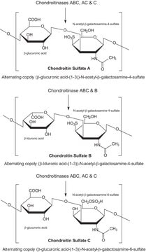
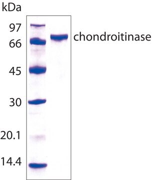
Chondroitinase ABC catalyzes the eliminative degradation of polysaccharides containing (1-4)-β-D-hexosaminyl and (1-3)-β-D-glucuronosyl or (1-3)-α-L-iduronosyl linkages to disaccharides containing 4-deoxy-β-D-gluc-4-enuronosyl groups. It acts on chondroitin 4-sulfate, chondroitin 6-sulfate, and dermatan sulfate, and acts slowly on hyaluronate. The molecular weight is found to be approximately 120 kDa with 2 non-identical subunits of molecular masses 86 kDa and 32 kDa. The pH optimum is found to be 8.0 with chondroitin sulfate and 6.8 with hyaluronic acid and temperature optimum is 37 °C. 1 mM Zn2+ acts as an inhibitor, while 0.05 M acetate is an activator of the enzyme.

Packaging

Packages based on chondroitinase C activity.

Other Notes

Contains potassium phosphate buffer salts and stabilizer

Unit Definition

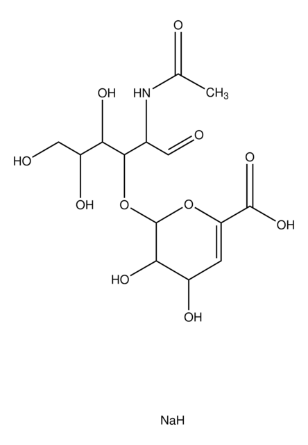
One unit will liberate 1.0 μmole of a mixture of 2-acetamido-2-deoxy-3-O-(β-D-gluc-4-ene-pyranosyluronic acid)-4-O-sulfo-D-galactose and 1.0 μmole of 2-acetamido-2-deoxy-3-O-(β-D-gluc-4-ene-pyranosyluronic acid)-6-O-sulfo-D-galactose from chondroitin sulfate from shark cartilage, per min at pH 8.0 at 37 °C.

Preparation Note

Affinity purified

Storage Class Code

11 - Combustible Solids

WGK

WGK 3

Flash Point(F)

Not applicable

Flash Point(C)

Not applicable

Personal Protective Equipment

dust mask type N95 (US), Eyeshields, Gloves

Certificates of Analysis (COA)

Search for Certificates of Analysis (COA) by entering the products Lot/Batch Number. Lot and Batch Numbers can be found on a product’s label following the words ‘Lot’ or ‘Batch’.

Already Own This Product?

Find documentation for the products that you have recently purchased in the Document Library.

Visit the Document Library

G Brückner et al.
Experimental brain research, 121(3), 300-310 (1998-09-24)
Lattice-like perineuronal accumulations of extracellular-matrix proteoglycans have been shown to develop during postnatal maturation and to persist throughout life as perineuronal nets (PNs) in many brain regions. However, the dynamics of their reorganization in adults are as yet unknown. The
Elizabeth J Bradbury et al.
Nature, 416(6881), 636-640 (2002-04-12)
The inability of axons to regenerate after a spinal cord injury in the adult mammalian central nervous system (CNS) can lead to permanent paralysis. At sites of CNS injury, a glial scar develops, containing extracellular matrix molecules including chondroitin sulphate
Rong-Rong Zhao et al.
Neuroscience bulletin, 29(4), 477-483 (2013-07-11)
After spinal cord injury (SCI), re-establishing functional circuitry in the damaged central nervous system (CNS) faces multiple challenges including lost tissue volume, insufficient intrinsic growth capacity of adult neurons, and the inhibitory environment in the damaged CNS. Several treatment strategies
P N Bansal et al.
Osteoarthritis and cartilage, 18(2), 184-191 (2009-10-10)
An early hallmark of osteoarthritis (OA) is the progressive loss of glycosaminoglycans (GAGs), the extracellular matrix (ECM) component of articular cartilage that confers it with compressive stiffness. Our aim in this work is to establish the feasibility of using Contrast
Susan M Smith et al.
International journal of molecular sciences, 20(3) (2019-01-27)
The aim of this study was to assess if the ovine articular cartilage serine proteinase inhibitors (SPIs) were related to the Kunitz inter-α-trypsin inhibitor (ITI) family. Ovine articular cartilage was finely diced and extracted in 6 M urea and SPIs

Articles

Uncover more about glycosaminoglycans and proteoglycans including the structure of glycosaminoglycans (GAGs), the different types of GAGs, and their functions.

Glycosaminoglycans are large linear polysaccharides constructed of repeating disaccharide units.

Our team of scientists has experience in all areas of research including Life Science, Material Science, Chemical Synthesis, Chromatography, Analytical and many others.

Contact Technical Service