CYTODET-RO
Roche
Cytotoxicity Detection KitPLUS (LDH)
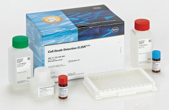
sufficient for 400 multiwell tests (04744926001), sufficient for 2,000 multiwell tests (04744934001), kit of 1 (4 components), suitable for protein quantification, suitable for cell analysis, detection
About This Item
Recommended Products
form
liquid
Quality Level
usage
sufficient for 2,000 multiwell tests (04744934001)
sufficient for 400 multiwell tests (04744926001)
packaging
kit of 1 (4 components)
manufacturer/tradename
Roche
storage condition
avoid repeated freeze/thaw cycles
technique(s)
protein quantification: suitable
λmax
490-492 nm
application(s)
cell analysis
detection
detection method
colorimetric
storage temp.
−20°C
General description
Application
- The Cytotoxicity Detection KitPLUS (LDH) is a fast, sensitive, and simple method to quantify cytotoxicity/cytolysis based on the measurement of LDH activity released from damaged cells using the 96-well or 384-well plate format. The kit can be used in many different in vitro cell systems when damage to the plasma membrane occurs. For example: Detection and quantification of cell-mediated cytotoxicity.
- Determination of mediator-induced cytolysis.
- Determination of the cytotoxic potential of compounds in environmental and medical research, and in the food, cosmetic, and pharmaceutical industries.
- Determination of cell death in bioreactors.
Features and Benefits
- Flexible, since the assay can determine the degree of plasma membrane damage in many different in vitro cell systems.
- Safe, because the assay does not use radioactive isotopes.
- Accurate, since assay results strongly correlate to the number of lysed cells.
- Sensitive, since the assay can detect even low cell numbers.
- Fast, since assay results can be determined on a multi-well ELISA reader, which allows simultaneous processing of many samples.
- Convenient, since the assay does not require a prelabeling step.
Packaging
- 04744926001 (1 kit containing 4 components)
- 04744934001 (1 kit containing 4 components)
Preparation Note
Dissolve the lyophilizate in 1 ml double dist. water for 10 min, then mix thoroughly.
Reaction mixture:
For 100 tests: Shortly before use, mix 250 μl of reconstituted catalyst with 11.25 ml dye solution
For 400 tests: Shortly before use, add the total volume of reconstituted catalyst (1 ml) to the total volume of dye solution (45 ml) and mix well.
Note: In case you observe crystals in “Dye solution”, shake the bottle at least 1 hour at 37 °C. Remaining precipitates will NOT influence the performance.
Note: The precipitates were formed during the freezing of the product. Repeated freezing and thawing should be avoided.
Note: Prepare immediately before use. Do not store the Reaction mixture.
Storage conditions (working solution): The lyophilizate is stable at 2 to 8 °C.
The reconstituted catalyst solution, the dye solution the lysis solution and the stop solution are stable for
- Four weeks when stored at 2 to 8 °C
- Two days at 15 to 25 °C
- For long-term storage (up to 3 months), store bottles at -15 to -25 °C.
- Freezing and thawing the solutions up to three times will not significantly reduce their performance. When crystals are seen in Dye solution, shake the bottle at least one hour at 37 °C. Remaining precipitates will not influence performance. As the precipitates can be formed during freezing of the product, repeated freezing and thawing should be avoided.
Other Notes
Kit Components Only
- Catalyst (Diaphorase/NAD+ mixture)
- Dye Solution (INT and sodium lactate)
- Lysis Solution
- Stop Solution
related product
Signal Word
Danger
Hazard Statements
Precautionary Statements
Hazard Classifications
Aquatic Chronic 3 - Eye Dam. 1 - Met. Corr. 1
Storage Class Code
8B - Non-combustible, corrosive hazardous materials
WGK
WGK 2
Flash Point(F)
does not flash
Flash Point(C)
does not flash
Certificates of Analysis (COA)
Search for Certificates of Analysis (COA) by entering the products Lot/Batch Number. Lot and Batch Numbers can be found on a product’s label following the words ‘Lot’ or ‘Batch’.
Already Own This Product?
Find documentation for the products that you have recently purchased in the Document Library.
Customers Also Viewed
Articles
Quality control guidelines to maintain high quality authenticated and contamination-free cell cultures. Free ECACC handbook download.
Our team of scientists has experience in all areas of research including Life Science, Material Science, Chemical Synthesis, Chromatography, Analytical and many others.
Contact Technical Service

