AB9018
Anti-Muscarinic Acetylcholine Receptor m3 Antibody
Chemicon®, from rabbit
Sign Into View Organizational & Contract Pricing
All Photos(1)
About This Item
UNSPSC Code:
12352203
eCl@ss:
32160702
NACRES:
NA.41
Recommended Products
biological source
rabbit
Quality Level
antibody form
affinity purified immunoglobulin
antibody product type
primary antibodies
clone
polyclonal
purified by
affinity chromatography
species reactivity
rat
manufacturer/tradename
Chemicon®
technique(s)
western blot: suitable
NCBI accession no.
UniProt accession no.
shipped in
wet ice
target post-translational modification
unmodified
Gene Information
rat ... Chrm3(24260)
Specificity
Muscarinic acetylcholine receptor m3.
Immunogen
Synthetic peptide from the 3rd intracellular loop of rat m3 (Accession P08483). The immunogen sequence is identical in human, mouse, bovine and porcine.
Application
Research Category
Neuroscience
Neuroscience
Research Sub Category
Neurotransmitters & Receptors
Neurotransmitters & Receptors
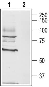
This Anti-Muscarinic Acetylcholine Receptor m3 Antibody is validated for use in WB for the detection of Muscarinic Acetylcholine Receptor m3.
Western blot: 1:2000 using ECL on rat brain lysate.
Dilutions should be made using a carrier protein such as BSA (1-3%)
Optimal working dilutions must be determined by the end user.
SUGGESTED WESTERN BLOT PROTOCOL
1. Mix the samples (organ membranes: 50 μg/lane; transfected cells: 500,000 cells/lane) with sample-buffer X 2, and heat 10 min at 70°C.
2. 5-50 μL applied to Minigel lane (0.75-1.5 mm width) and run at standard conditions. (60 mA for 2 1.5 mm Minigel gels, 1.4 h). It is suggested that you run 5-15% acrylamide (37.5:1 acrylamide:bisacrysmide) minigel (1.5 mm width) at 30 mA/gel ~1-1.5 hours.
3. Transfer in semi-dry system under standard conditions (3 h 100 mA for two minigel gels)
4. Stain the transferred bands with Chemicon BLOT-FastStain (Catalog Number 2076).
5. Destain with deionized water.
6. Block with 5% non-fat milk (Marvel or Carnation) in PBS, and 0.025 % sodium azide, overnight at 2-8°C. The non-fat milk should be dissolved freshly, centrifuged 10,000 rpm for 10 min, and filtered through glass filter (Gelman Acrodisc).
7. Incubation with first antibody 2 h at room temperature or overnight at 4°C in blocking solution. The antibody preparation should be centrifuged before use (10,000 g 5 min.). Optimal working dilutions and incubation time will need to be determined by the end user.
8. Wash 4 x 10 min. with PBS-0.1% tween 20. From this stage, azide should be omitted.
9. Incubation with the secondary antibody (HRP-conjugated goat anti-rabbit antibody, for example Chemicon Catalog Number AP132P, diluted appropriately) 1 h at room temperature.
10. Wash 4 x 10 min. with PBS-0.1% tween 20.
11. Perform ECL with commercial kits (Chemilucent, Chemicon Catalog Number 2600).
Physical form
Affinity purified immunoglobulin. Lyophilized from phosphate buffered saline, pH 7.4, containing 1% BSA, and 0.05% sodium azide as a preservative. Reconstitute with 50 μL of sterile deionized water. Centrifuge antibody preparation before use (10,000 xg for 5 min).
Storage and Stability
Maintain lyophilized material at -20°C for up to 12 months after date of receipt. After reconstitution maintain at -20°C in undiluted aliquots for up to 6 months. Avoid repeated freeze/thaw cycles.
Analysis Note
Control
Included free of charge with the antibody is 40 μg of control antigen. The stock solution of the antigen can be made up using 100 μL of sterile distilled water. For negative control, preincubate 1 μg of peptide with 1 μg of antibody for one hour at room temperature. Optimal concentrations must be determined by the end user.
Included free of charge with the antibody is 40 μg of control antigen. The stock solution of the antigen can be made up using 100 μL of sterile distilled water. For negative control, preincubate 1 μg of peptide with 1 μg of antibody for one hour at room temperature. Optimal concentrations must be determined by the end user.
Other Notes
Concentration: Please refer to the Certificate of Analysis for the lot-specific concentration.
Legal Information
CHEMICON is a registered trademark of Merck KGaA, Darmstadt, Germany
Disclaimer
Unless otherwise stated in our catalog or other company documentation accompanying the product(s), our products are intended for research use only and are not to be used for any other purpose, which includes but is not limited to, unauthorized commercial uses, in vitro diagnostic uses, ex vivo or in vivo therapeutic uses or any type of consumption or application to humans or animals.
Not finding the right product?
Try our Product Selector Tool.
Hazard Statements
Precautionary Statements
Hazard Classifications
Aquatic Chronic 3
Storage Class Code
11 - Combustible Solids
WGK
WGK 3
Certificates of Analysis (COA)
Search for Certificates of Analysis (COA) by entering the products Lot/Batch Number. Lot and Batch Numbers can be found on a product’s label following the words ‘Lot’ or ‘Batch’.
Already Own This Product?
Find documentation for the products that you have recently purchased in the Document Library.
Michael Winder et al.
Basic & clinical pharmacology & toxicology, 121(4), 257-265 (2017-04-25)
In the urinary bladder, the main source of NO seems to be the urothelium and the underlying suburothelium. In this study, we aimed to characterize how receptors in the human urothelium regulate the production and release of NO. For this
Masato Asahina et al.
Internal medicine (Tokyo, Japan), 52(24), 2733-2737 (2013-12-18)
The autoimmune mechanism is considered to play an important role in the development of acquired idiopathic generalized anhidrosis (AIGA), and muscarinic M3 receptors (M3Rs) on eccrine glands are possible autoimmune targets. We investigated the existence of autoantibodies against M3Rs in
Marcy L Guerra et al.
The Journal of biological chemistry, 289(20), 14370-14379 (2014-04-04)
We have shown recently that the class C G protein-coupled receptor T1R1/T1R3 taste receptor complex is an early amino acid sensor in MIN6 pancreatic β cells. Amino acids are unable to activate ERK1/2 in β cells in which T1R3 has
Martin Dankis et al.
Investigative ophthalmology & visual science, 62(12), 19-19 (2021-09-22)
The functional characteristics of receptors that regulate lacrimal gland myoepithelial cells are still somewhat unclear. To date, mainly muscarinic receptors have been of interest; however, further knowledge is needed regarding their expression and functional roles. For this purpose, primary cultures
Daniel Giglio et al.
The Journal of pharmacology and experimental therapeutics, 365(2), 327-335 (2018-03-14)
Currently, we have assessed the neuronal control of the urinary bladder in radiation cystitis and whether interstitial cells contribute to the condition. Fourteen days after bladder irradiation (20 Gy), rats were sedated and the urinary bladder was cut into two
Our team of scientists has experience in all areas of research including Life Science, Material Science, Chemical Synthesis, Chromatography, Analytical and many others.
Contact Technical Service


