1.06887
Papanicolaou′s solution 2b Orange II solution
for cytology
About This Item
Recommended Products
Quality Level
form
liquid
IVD
for in vitro diagnostic use
pH
7.0-7.5 (20 °C in H2O)
transition temp
flash point 17 °C
density
0.83 g/cm3 at 20 °C
application(s)
clinical testing
diagnostic assay manufacturing
hematology
histology
storage temp.
15-25°C
Related Categories
General description
In the first step, the cell nuclei are stained either progressively or regressively with a hematoxylin solution, (Hematoxylin solution modified acc.to Gill II, (Product number 1.05175),
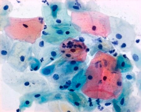
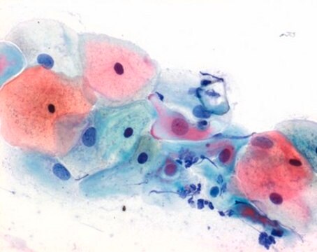
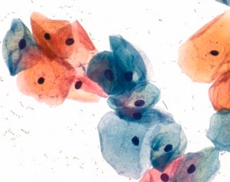
Papanicolaou′s solution 1a Harris hematoxylin solution, (Product number 1.09253) or Papanicolaou′s solution 1b Hematoxylin solution S, (Product number 1.09254). Nuclei are stained blue to dark violet. In the progressive hematoxylin staining method, staining is carried out to the endpoint, after which the slide is blued in tapwater. With the regressive method the material is over-stained and the excess of staining solution is removed by acid rinsing steps, followed by the bluing step. The structures of nuclei are more differentiated and better visible by the regressive method. The second staining step is cytoplasmic staining by orange staining solution, (Papanicolaou′s solution 2b Orange II solution, (Product number 1.106887) or Papanicolaou′s solution 2a Orange G solution (OG6), (Product number 1.06888)), especially for demonstration of mature and keratinized cells. The target structures are stained orange in different intensities. In the third staining step is used the so-called polychrome solution, a mixture of eosin, light green SF and Bismarck brown, Papanicolaou′s solution 3a polychromatic solution EA 31, (Product number 1.09271), Papanicolaou′s solution 3b polychromatic solution EA 50, (Product number 1.09272), Papanicolaou′s solution 3c polychromatic solution EA 65, (Product number 1.09270), or Papanicolaou′s solution 3d polychromatic solution EA 65, (Product number 1.09269). The polychrome solution is used for demonstration of differentiation of squamous cells. Papanicolaou′s solution 2b Orange II solution gives a more intense reddish staining result with mature and keratinized squamous cells than Papanicolaou′s solution 2a Orange G solution (OG6) - for cytology.
The 500 ml bottle provides 1500 - 2000 stainings. For more details, please see instructions for use (IFU). This product is registered as IVD and CE marked.
Analysis Note
Nuclei: blue to dark violet
Cyanophilic cytoplasm: blue-green
Eosinophilic cytoplasm: pink
signalword
Danger
Hazard Classifications
Acute Tox. 4 Dermal - Acute Tox. 4 Inhalation - Acute Tox. 4 Oral - Eye Irrit. 2 - Flam. Liq. 2 - STOT SE 1
Storage Class
3 - Flammable liquids
wgk_germany
WGK 2
flash_point_f
62.6 °F
flash_point_c
17 °C
Certificates of Analysis (COA)
Search for Certificates of Analysis (COA) by entering the products Lot/Batch Number. Lot and Batch Numbers can be found on a product’s label following the words ‘Lot’ or ‘Batch’.
Already Own This Product?
Find documentation for the products that you have recently purchased in the Document Library.
Our team of scientists has experience in all areas of research including Life Science, Material Science, Chemical Synthesis, Chromatography, Analytical and many others.
Contact Technical Service


