Skip to Content
MilliporeSigma
All Photos(1)

Key Documents

07-2268

Sigma-Aldrich

Anti-phospho-TBC1D1 Antibody (Ser237)

from rabbit, purified by affinity chromatography

Synonym(s):

TBC1 domain family member 1, TBC1 (tre-2/USP6, BUB2, cdc16) domain family, member 1

Sign Into View Organizational & Contract Pricing


About This Item

UNSPSC Code:
12352203
eCl@ss:
32160702

biological source

rabbit

Quality Level

antibody product type

primary antibodies

clone

polyclonal

purified by

affinity chromatography

species reactivity

human

species reactivity (predicted by homology)

ox (based on 100% sequence homology), chimpanzee (based on 100% sequence homology), mouse (based on 100% sequence homology), rat (based on 100% sequence homology), rhesus macaque (based on 100% sequence homology)

technique(s)

western blot: suitable

NCBI accession no.

UniProt accession no.

shipped in

wet ice

target post-translational modification

phosphorylation (pSer237)

Gene Information

human ... TBC1D1(23216)

General description

TBC1D1 is the first member of a family of proteins that contain the tbc box motif of 180-220 amino acids and may be related to cell growth and differentiation. The composition of TBC1D1 has the same function as a known regulator of insulin-mediated Glut4 translocation and may play a role in severe human obesity. An insulin-independent increase in glucose transport several hours after exercise has been attributed to TBC1D1 phosphorylation. The R125W mutation of TBC1D1 is thought to decrease skeletal muscle glucose transport and could be the main reason for the obesity that seems to be linked to this type of mutation.

Specificity

This antibody recognizes TBC1D1 phosphorylated at Ser237.

Immunogen

Epitope: Phosphorylated Ser237
KLH-conjugated linear peptide corresponding to human TBC1D1 phosphorylated at Ser237.

Application

Research Category
Signaling
Research Sub Category
Insulin/Energy Signaling

Glucose/Glycogen Metabolism
This phospho-TBC1D1 antibody is validated for use in WB for the detection of the phospho-TBC1D1 protein.

Quality

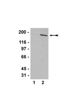
Evaluated by Western Blot in lambda phosphatase treated and untreated HEK293 cell lysates.

Western Blot Analysis: 1 µg/mL of this antibody detected TBC1D1 on 10 µg of lambda phosphatase treated and untreated HEK293 cell lysates.

Target description

~145 kDa observed

Physical form

Affinity purified
Purified rabbit polyclonal in buffer containing 0.1 M Tris-Glycine (pH 7.4), 150 mM NaCl with 0.05% sodium azide.

Storage and Stability

Stable for 1 year at 2-8°C from date of receipt.

Analysis Note

Control
Lambda phosphatase treated and untreated HEK293 cell lysates

Other Notes

Concentration: Please refer to the Certificate of Analysis for the lot-specific concentration.

Disclaimer

Unless otherwise stated in our catalog or other company documentation accompanying the product(s), our products are intended for research use only and are not to be used for any other purpose, which includes but is not limited to, unauthorized commercial uses, in vitro diagnostic uses, ex vivo or in vivo therapeutic uses or any type of consumption or application to humans or animals.

Not finding the right product?  

Try our Product Selector Tool.

Storage Class Code

12 - Non Combustible Liquids

WGK

WGK 1

Flash Point(F)

Not applicable

Flash Point(C)

Not applicable


Certificates of Analysis (COA)

Search for Certificates of Analysis (COA) by entering the products Lot/Batch Number. Lot and Batch Numbers can be found on a product’s label following the words ‘Lot’ or ‘Batch’.

Already Own This Product?

Find documentation for the products that you have recently purchased in the Document Library.

Visit the Document Library

Marie Locard-Paulet et al.
Molecular systems biology, 16(7), e9524-e9524 (2020-07-04)
T-cell receptor (TCR) ligation-mediated protein phosphorylation regulates the activation, cellular responses, and fates of T cells. Here, we used time-resolved high-resolution phosphoproteomics to identify, quantify, and characterize the phosphorylation dynamics of thousands of phosphorylation sites in primary T cells during
Sharon C Hook et al.
Scientific reports, 10(1), 17953-17953 (2020-10-23)
Proteins involved in the spaciotemporal regulation of GLUT4 trafficking represent potential therapeutic targets for the treatment of insulin resistance and type 2 diabetes. A key regulator of insulin- and exercise-stimulated glucose uptake and GLUT4 trafficking is TBC1D1. This study aimed
Jonas Roland Knudsen et al.
Experimental physiology, 104(5), 704-714 (2019-02-03)
What is the central question of this study? Resolving the mechanism(s) leading to glucose transporter 4 (GLUT4) translocation to the muscle surface membrane has great therapeutic potential. However, the measurement of GLUT4 translocation is technically challenging. Here, we asked whether
Yang Liu et al.
Nature, 578(7796), 605-609 (2020-02-14)
The activation of adenosine monophosphate-activated protein kinase (AMPK) in skeletal muscle coordinates systemic metabolic responses to exercise1. Autophagy-a lysosomal degradation pathway that maintains cellular homeostasis2-is upregulated during exercise, and a core autophagy protein, beclin 1, is required for AMPK activation in

Our team of scientists has experience in all areas of research including Life Science, Material Science, Chemical Synthesis, Chromatography, Analytical and many others.

Contact Technical Service