Skip to Content
MilliporeSigma
All Photos(2)

Key Documents

ABS811

Sigma-Aldrich

Anti-FABP7 Antibody

from rabbit, purified by affinity chromatography

Synonym(s):

Fatty acid-binding protein, brain, Brain lipid-binding protein, BLBP, Brain-type fatty acid-binding protein, B-FABP, Fatty acid-binding protein 7, Mammary-derived growth inhibitor related, FABP7

Sign Into View Organizational & Contract Pricing


About This Item

UNSPSC Code:
12352203
eCl@ss:
32160702
NACRES:
NA.41

biological source

rabbit

Quality Level

antibody form

affinity isolated antibody

antibody product type

primary antibodies

clone

polyclonal

purified by

affinity chromatography

species reactivity

human

technique(s)

immunohistochemistry: suitable
western blot: suitable

UniProt accession no.

shipped in

wet ice

target post-translational modification

unmodified

Gene Information

human ... FABP7(2173)

General description

FABP7 is a member of the large family of hydrophobic ligand-binding proteins (FABPs). FABP proteins are involved in lipid metabolism, including uptake and intracellular trafficking of fatty acids and retinoids. In addition, they play a role in gene regulation, cell signaling, cell growth and differentiation. Several reports have suggested a possible role for the different FABP proteins in cancer biology, linking their levels with either increasing or decreasing the degree of malignancy. Nine FABPs (FABP1-FABP9) have been identified. FABP7 is a brain-specific protein that is required for the establishment of the radial glial fiber system in the developing brain, a system that is necessary for the migration of immature neurons to establish cortical layers. It is expressed at high levels in the developing central nervous system. FABP7 has also been found in melanocytic lesions and has shown to regulate proliferation and invasion in melanoma cells in vitro, suggesting that it may play a role in cell proliferation and invasion.

Immunogen

Epitope: Near Center
FABP7 antibody was raised against a 17 amino acid synthetic peptide from near the center of human FABP7.

Application

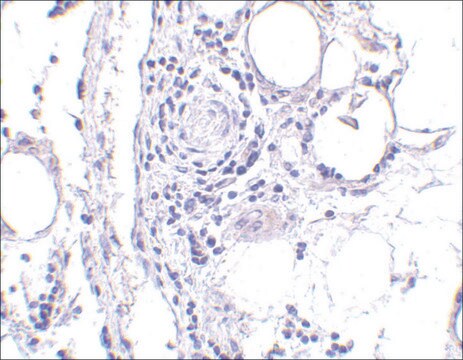
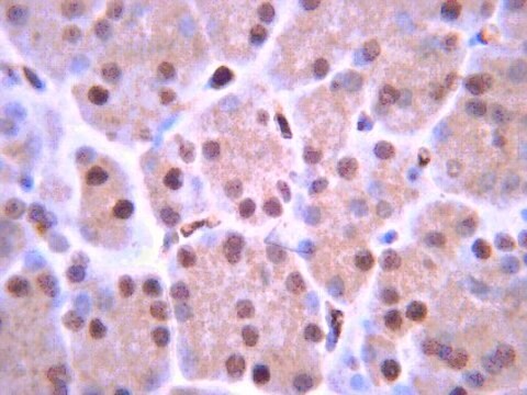
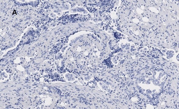
Immunohistochemistry Analysis (IHC): 5 μg/mL from a representative lot detected FABP7 in human breast tissue.

Research Category
Signaling
Research Sub Category
Developmental Signaling
This Anti-FABP7 Antibody is validated for use in Western Blotting_x000D_ and Immunohistochemistry for the detection of FABP7.

Quality

Evaluated by Western Blotting in human breast tissue lysate.

Western Blotting Analysis (WB): 1 μg/mL of this antibody detected FABP7 in human breast tissue lysate.

Target description

~24 kDa observed. Uncharacterized bands may be observed in some lysate(s).

Linkage

Replaces: MABS184

Physical form

Antigen Affinity Purified
Purified rabbit polyclonal in buffer containing PBS (pH 7.2) with 0.02% sodium azide.

Storage and Stability

Stable for 1 year at 2-8°C from date of receipt.

Other Notes

Concentration: Please refer to lot specific datasheet.

Disclaimer

Unless otherwise stated in our catalog or other company documentation accompanying the product(s), our products are intended for research use only and are not to be used for any other purpose, which includes but is not limited to, unauthorized commercial uses, in vitro diagnostic uses, ex vivo or in vivo therapeutic uses or any type of consumption or application to humans or animals.

Not finding the right product?  

Try our Product Selector Tool.

Storage Class Code

12 - Non Combustible Liquids

WGK

WGK 2

Flash Point(F)

Not applicable

Flash Point(C)

Not applicable


Certificates of Analysis (COA)

Search for Certificates of Analysis (COA) by entering the products Lot/Batch Number. Lot and Batch Numbers can be found on a product’s label following the words ‘Lot’ or ‘Batch’.

Already Own This Product?

Find documentation for the products that you have recently purchased in the Document Library.

Visit the Document Library

Nolwenn Pasquet et al.
Cerebral cortex (New York, N.Y. : 1991), 29(6), 2482-2498 (2018-06-08)
Modifications of neuronal migration during development, including processes that control cortical lamination are associated with functional deficits at adult stage. Here, we report for the first time that the lack of the serine protease tissue-type Plasminogen Activator (tPA), previously characterized
Brian G Rash et al.
The Journal of neuroscience : the official journal of the Society for Neuroscience, 31(43), 15604-15617 (2011-10-28)
The processes regulating cortical surface area expansion during development and evolution are unknown. We show that loss of function of all fibroblast growth factor receptors (FgfRs) expressed at the earliest stages of cortical development causes severe deficits in surface area

Our team of scientists has experience in all areas of research including Life Science, Material Science, Chemical Synthesis, Chromatography, Analytical and many others.

Contact Technical Service