Skip to Content
MilliporeSigma
All Photos(2)

Key Documents

MAB5382

Sigma-Aldrich

Anti-Hypoxia Inducible Factor 1 α Antibody, clone H1α67

clone H1alpha67, Chemicon®, from mouse

Synonym(s):

HIF-1 alpha, ARNT Interacting Protein, MOP1

Sign Into View Organizational & Contract Pricing


About This Item

UNSPSC Code:
12352203
eCl@ss:
32160702
NACRES:
NA.41

biological source

mouse

Quality Level

antibody form

purified immunoglobulin

antibody product type

primary antibodies

clone

H1alpha67, monoclonal

species reactivity

mouse, rat, ferret, human

packaging

antibody small pack of 25 μg

manufacturer/tradename

Chemicon®

technique(s)

electrophoretic mobility shift assay: suitable
immunohistochemistry: suitable
immunoprecipitation (IP): suitable
western blot: suitable

isotype

IgG2b

NCBI accession no.

UniProt accession no.

shipped in

ambient

storage temp.

2-8°C

target post-translational modification

unmodified

Gene Information

human ... HIF1A(3091)

General description

HIF is a transcriptional complex that plays a central role in mammalian oxygen homeostasis, the posttranslational modification by prolyl hydroxylation as a key regulatory event that targets HIF-alpha subunits for proteasomal destruction via the von Hippel-Lindau ubiquitylation complex. The transcriptional complex is composed of an alpha-beta heterodimer; HIF-beta being a constitutive nuclear protein that dimerises with oxygen regulated HIF-alpha subunits. In normoxia, 4-hydroxylation of human HIF-alpha at Pro402 or Pro564 by a set of HIF prolyl hydroxylase isoenzymes (PHD 1-3) mediates HIF1-alpha recognition by von Hippel-Lindau ubiquitin ligase complex leading to its proteasomal destruction. In hypoxia (deprivation of oxygen), lack of hydroxylase activity enables HIF-alpha subunits to escape destruction and become transcriptionally active. Thus HIF hydroxylases provide a focus for understanding cellular responses to hypoxia and target for therapeutic manipulation. There are several HIF factors, which include HIF 1-alpha, HIF 1-beta, HIF 2-alpha. HIF 1-alpha is an 812 a.a. protein in rat and 836 a.a. long in mouse and human. A master transcriptional regulator of the adaptive response to hypoxia. Under hypoxic conditions activates the transcription of over 40 genes, including, erythropoietin, glucose transporters, glycolytic enzymes, vascular endothelial growth factor, and other genes whose protein products increase oxygen delivery or facilitate metabolic adaptation to hypoxia. Plays an essential role in embryonic vascularization, tumor angiogenesis and pathophysiology of ischemic disease. It is ubiquitous in expression as cytoplasmic in normoxia, nuclear translocation in response to hypoxia.

Specificity

Recognizes Hypoxia inducible factor-1alpha (HIF-1alpha).

Immunogen

Fusion protein from amino acids 432-528 of human HIF-1alpha.

Application

Anti-Hypoxia Inducible Factor 1 α Antibody, clone H1α67 is a Mouse Monoclonal Antibody for detection of Hypoxia Inducible Factor 1 α also known as HIF-1 alpha or ARNT Interacting Protein & has been validated in EMSA, IHC, IP & WB.
Research Category
Epigenetics & Nuclear Function
Research Sub Category
Transcription Factors
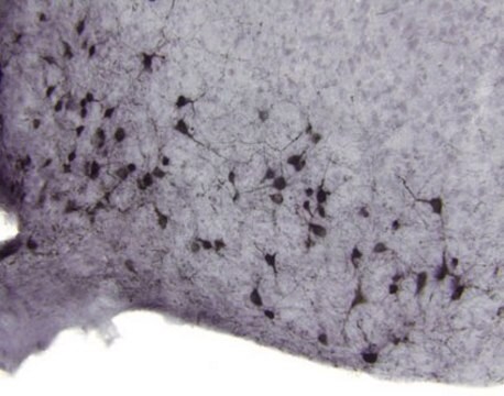
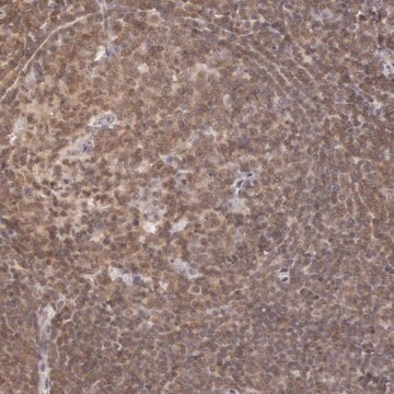
Western blot: 1:500-1:1,000. The antibody recognizes a band of 120 kD in induced tissues and cells. Multiple bands may be present at 120 kD representing post-translational modification of HIF-1alpha.

Immunohistochemistry: 1:500-1:1,000. The antibody has been used successfully on formalin-fixed, paraffin embedded tissue sections after antigen retrieval.

Immunoprecipitation

Gel Shift

Optimal working dilutions must be determined by end user.

Target description

Predicted MW: 96 kDa, apparent MW: 120 kDa

Linkage

Replaces: 04-1006

Physical form

Format: Purified
Protein A Purified mouse immunoglobulin in 20 mM sodium phosphate, 250 mM NaCl, pH. 7.6, with 0.1% sodium azide as a preservative.
Protein A purified

Storage and Stability

Maintain for 1 year at 2–8°C from date of shipment. Aliquot to avoid repeated freezing and thawing. For maximum recovery of product, centrifuge the original vial after thawing and prior to removing the cap.

Analysis Note

Control
Cobalt chloride-treated MCF-7 cells

Other Notes

Concentration: Please refer to the Certificate of Analysis for the lot-specific concentration.

Legal Information

CHEMICON is a registered trademark of Merck KGaA, Darmstadt, Germany

Disclaimer

Unless otherwise stated in our catalog or other company documentation accompanying the product(s), our products are intended for research use only and are not to be used for any other purpose, which includes but is not limited to, unauthorized commercial uses, in vitro diagnostic uses, ex vivo or in vivo therapeutic uses or any type of consumption or application to humans or animals.

Not finding the right product?  

Try our Product Selector Tool.


Certificates of Analysis (COA)

Search for Certificates of Analysis (COA) by entering the products Lot/Batch Number. Lot and Batch Numbers can be found on a product’s label following the words ‘Lot’ or ‘Batch’.

Already Own This Product?

Find documentation for the products that you have recently purchased in the Document Library.

Visit the Document Library

Hiroko Seino et al.
Molecular medicine reports, 13(5), 3821-3827 (2016-04-02)
Several studies have noted benign thecoma‑fibroma tumors with positive F‑18 fluorodeoxyglucose (FDG) accumulation mimicking malignant ovarian tumors following F‑18 FDG positron emission tomography (PET). The present study analyzed four cases with false‑positive F‑18 FDG PET/computed tomography (CT) diagnoses of thecoma‑fibroma
Bioimaging assessment and effect of skin wound healing using bone-marrow-derived mesenchymal stromal cells with the artificial dermis in diabetic rats.
Hirokazu Inoue, Takashi Murakami, Takashi Ajiki, Mayumi Hara, Yuichi Hoshino et al.
Journal of Biomedical Optics null
Hadas Pahima et al.
Oncotarget, 9(16), 12825-12841 (2018-03-22)
The voltage-dependent anion channel 1 (VDAC1), an outer mitochondria membrane (OMM) protein, serves as a mitochondrial gatekeeper, mediating the transport of nucleotides, Ca2+ and other metabolites across the OMM. VDAC1 also plays a central role in mitochondria-mediated apoptosis by facilitating
[Abstracts of the 95th Annual Meeting of the German Society of Pathology. June 16-19, 2011. Leipzig, Germany].
Der Pathologe, 32 Suppl 1, 5-147 (2011-05-06)
Limin Gu et al.
Eye (London, England), 33(4), 600-609 (2018-11-08)
Diabetes causes various biochemical changes in the retina; long-term changes in the factors associated with hypoxia and gliosis have rarely been reported. The present study was conducted to explore the changes in these factors in a time-dependent manner in experimental

Our team of scientists has experience in all areas of research including Life Science, Material Science, Chemical Synthesis, Chromatography, Analytical and many others.

Contact Technical Service