Skip to Content
MilliporeSigma
All Photos(4)

Key Documents

MABC1108

Sigma-Aldrich

Anti-LAMP-1 Antibody, clone H4A3

clone H4A3, from mouse

Synonym(s):

Lysosome-associated membrane glycoprotein 1, Lysosome-associated membrane protein 1, CD107 antigen-like family member A, CD107a

Sign Into View Organizational & Contract Pricing


About This Item

UNSPSC Code:
12352203
eCl@ss:
32160702
NACRES:
NA.43

biological source

mouse

antibody form

purified immunoglobulin

antibody product type

primary antibodies

clone

H4A3, monoclonal

species reactivity

human

packaging

antibody small pack of 25 μg

technique(s)

dot blot: suitable
immunofluorescence: suitable
immunohistochemistry: suitable (paraffin)
immunoprecipitation (IP): suitable
western blot: suitable

isotype

IgGκ

NCBI accession no.

UniProt accession no.

target post-translational modification

unmodified

Gene Information

human ... LAMP1(3916)

General description

Lysosome-associated membrane glycoprotein 1 (UniProt: P11279; also known as LAMP-1, Lysosome-associated membrane protein 1, CD107 antigen-like family member A, CD107a) is encoded by the LAMP1 gene (Gene ID: 3916) in human. LAMP-1 and LAMP-2 are two highly glycosylated proteins found in the lysosomal membrane. They may contribute up to 50% of all proteins present in the lysosomal membranes. They are known to maintain the lysosomal acidification and lysosomal membrane integrity. Extensive N-glycosylation on LAMP-1 and 2 to protects them from the lysosomal proteolysis. However, higher levels of poly-N-acetyllactosamines on LAMP-1 and 2 have been correlated with decreased cellular differentiation and enhanced metastatic potential. LAMP-1 is a single-pass type I transmembrane protein that contains a large lumenal domain (aa 29-382), a transmembrane domain (aa 383-405), and a short C-terminal cytoplasmic tail (aa 406-417). LAMP-1 is synthesized with a signal peptide (aa 1-28) that is subsequently cleaved in the mature form. LAMP-1 presents carbohydrate ligands to selectins and is also reported to act as a receptor for Lassa virus protein. Two isoforms of LAMP-1 have been described that are produced by alternative splicing. (Ref.: Tan, K-P., et al. (2016). Cell Death Dis. 7, e2347).

Specificity

Clone H4A3 detects Lysosome-associated membrane glycoprotein 1 (LAMP-1) in human cells.

Immunogen

Human adherent peripheral blood cells.

Application

Anti-LAMP-1, clone H4A3, Cat. No. MABC1108, is a mouse monoclonal antibody that detects Lysosome-associated membrane glycoprotein 1 (LAMP-1) and has been tested for use in Dot Blot, Immunofluorescence, Immunohistochemistry (Paraffin), Immunoprecipitation, and Western Blotting.
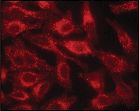
Immunocytochemistry Analysis: A 1:500 dilution from a representative lot detected LAMP-1 in HeLa and A431 cells.

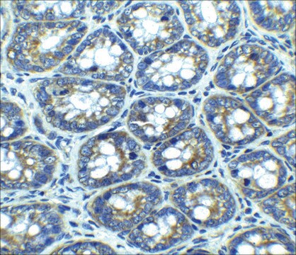
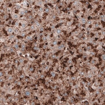
Immunohistochemistry (Paraffin) Analysis: A representative lot detected LAMP-1 in Immunohistochemistry applications (Furuta, K., et. al. (2001). Am J Pathol. 159(2):449-55).

Western Blotting Analysis: A representative lot detected LAMP-1 in Western Blotting applications (Mane, S.M., et. al. (1989). Arch Biochem Biophys. 268(1):360-78).

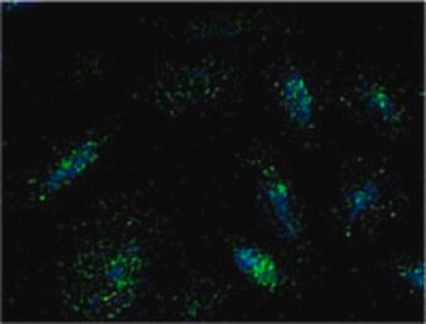
Immunofluorescence Analysis: A representative lot detected LAMP-1 in Immunofluorescence applications (Starr, T., et. al. (2011). Cell Host Microbe. 11(1):33-45).

Dot Blot Analysis: A representative lot detected LAMP-1 in Dot Blot applications (Arruda, L.B., et. al. (2006). J Immunol. 177(4):2265-75).

Immunohistochemistry (Paraffin) Analysis: A 1:250 dilution from a representative lot detected LAMP-1 in human kidney and human pancreas tissue sections.

Immunoprecipitation Analysis: A representative lot immunoprecipitated LAMP-1 in Immunoprecipitation applications (Mane, S.M., et. al. (1989). Arch Biochem Biophys. 268(1):360-78).
Research Category
Cell Structure

Quality

Evaluated by Western Blotting in HEK293 cell lysate.

Western Blotting Analysis: 1 µg/mL of this antibody detected LAMP-1 in HEK293 cell lysate.

Target description

~110 kDa observed; 44.88 kDa calculated. Uncharacterized bands may be observed in some lysate(s).

Physical form

Format: Purified
Protein G purified
Purified mouse monoclonal antibody IgG in buffer containing 0.1 M Tris-Glycine (pH 7.4), 150 mM NaCl with 0.05% sodium azide.

Storage and Stability

Stable for 1 year at 2-8°C from date of receipt.

Other Notes

Concentration: Please refer to lot specific datasheet.

Disclaimer

Unless otherwise stated in our catalog or other company documentation accompanying the product(s), our products are intended for research use only and are not to be used for any other purpose, which includes but is not limited to, unauthorized commercial uses, in vitro diagnostic uses, ex vivo or in vivo therapeutic uses or any type of consumption or application to humans or animals.

Not finding the right product?  

Try our Product Selector Tool.


Certificates of Analysis (COA)

Search for Certificates of Analysis (COA) by entering the products Lot/Batch Number. Lot and Batch Numbers can be found on a product’s label following the words ‘Lot’ or ‘Batch’.

Already Own This Product?

Find documentation for the products that you have recently purchased in the Document Library.

Visit the Document Library

Our team of scientists has experience in all areas of research including Life Science, Material Science, Chemical Synthesis, Chromatography, Analytical and many others.

Contact Technical Service