Skip to Content
MilliporeSigma
All Photos(2)

Key Documents

MABS1304

Sigma-Aldrich

Anti-ATP Synthase subunit β Antibody, clone 11/21-7-A8

clone 11/21-7-A8, from mouse

Synonym(s):

ATP synthase subunit beta, mitochondrial, ATP Synthase subunit β, beta-F1-ATPase

Sign Into View Organizational & Contract Pricing


About This Item

UNSPSC Code:
12352203
eCl@ss:
32160702
NACRES:
NA.41
clone:
11/21-7-A8, monoclonal
application:
DB
ELISA
ICC
WB
species reactivity:
human, mouse, rat
technique(s):
ELISA: suitable
dot blot: suitable
immunocytochemistry: suitable
western blot: suitable
citations:
3

biological source

mouse

Quality Level

antibody form

purified immunoglobulin

antibody product type

primary antibodies

clone

11/21-7-A8, monoclonal

species reactivity

human, mouse, rat

technique(s)

ELISA: suitable
dot blot: suitable
immunocytochemistry: suitable
western blot: suitable

isotype

IgG1κ

NCBI accession no.

UniProt accession no.

shipped in

wet ice

target post-translational modification

unmodified

Gene Information

human ... ATP5B(506)

General description

ATP synthase subunit beta, mitochondrial (EC 3.6.3.14; UniProt P06576; also known as ATP synthase H+ transporting mitochondrial F1 complex beta polypeptide, beta-F1-ATPase, Epididymis secretory protein Li 271, Mitochondrial ATP synthase beta subunit, Mitochondrial ATP synthetase beta subunit) is encoded by the ATP5B (also known as ATPMB, ATPSB, HEL-S-271) gene (Gene ID 506) in human. Mitochondrial ATP synthase produces ATP from ADP in the presence of a proton gradient generated by electron transport complexes. This ATPase contains two structural domains, F1-containing extramembrane catalytic core, and F0-containing the membrane proton channel that are linked via a central stalk and a peripheral stalk. Subunits alpha and beta form the catalytic code in F1. Tumor development requires the selection of cancer cells with a repressed biogenesis and functional activity of mitochondria. Both beta-F1-ATPase/GAPDH and beta-F1-ATPase/Hsp60 ratios are found to be significantly lower in tumors than the corresponding normal tissues. Studies conducted in human colon cancer cell line HCT116 show the involvement of AMPK (AMP-activated protein kinase) and GCN2 (general control non-derepressible 2; eIF2α kinase) in the onset of colon cancer progression by repressing of beta-F1-ATPase synthesis and promoting the abnormal bioenergetics of mitochondria.

Immunogen

His-tagged recombinant protein corresponding to human ATP Synthase subunit β.

Application

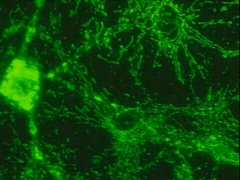
Immunocytochemistry Analysis: A representative lot immunostained mitochondrial tubular network in human breast cancer Hs578T cells (Acebo, P., et al (2009). Transl Oncol. 2(3):138-145).
ELISA Analysis: A representative lot detected His-tagged full-length human ATP Synthase subunit β (beta-F1-ATPase) recombinant protein by direct ELISA (Acebo, P., et al (2009). Transl Oncol. 2(3):138-145).
Dot Blot Analysis: A representative lot detected ATP Synthase subunit β (beta-F1-ATPase) by Dot blot using His-tagged full-length human beta-F1-ATPase recombinant protein or HepG2 lysate (Acebo, P., et al (2009). Transl Oncol. 2(3):138-145).
Western Blotting Analysis: A representative lot detected ATP Synthase subunit β (beta-F1-ATPase) in human hepatoma HepG2, murine hepatoma Hepa 1-6, and normal rat liver epithelial C9 (Clone 9) cells.
Western Blotting Analysis: A representative lot detected ATP Synthase subunit β (beta-F1-ATPase) expression in various cancer patients tissues (Acebo, P., et al (2009). Transl Oncol. 2(3):138-145).
Western Blotting Analysis: A representative lot detected ATP Synthase subunit β (beta-F1-ATPase) downregulation in HCT116 human colon cancer cells in response to AMPK pathway activation upon oligomycin or AICAR treatment (Martinez-Reyes, J., et al. (2012). Biochem J. 444(2):249-259).
Research Category
Signaling
Research Sub Category
Developmental Signaling
This Anti-ATP Synthase subunit β Antibody, clone 11/21-7-A8 is validated for use in Western Blotting, Immunocytochemistry, ELISA and Dot Blot for the detection of ATP Synthase subunit β.

Quality

Evaluated by Western Blotting in HepG2 cell lysate.

Western Blotting Analysis: 0.5 µg/mL of this antibody detected ATP Synthase subunit β in 10 µg of HepG2 cell lysate.

Target description

~56 kDa observed

Physical form

Format: Purified
Protein G Purified
Purified mouse monoclonal IgG1κ antibody in buffer containing 0.1 M Tris-Glycine (pH 7.4), 150 mM NaCl with 0.05% sodium azide.

Storage and Stability

Stable for 1 year at 2-8°C from date of receipt.

Other Notes

Concentration: Please refer to lot specific datasheet.

Disclaimer

Unless otherwise stated in our catalog or other company documentation accompanying the product(s), our products are intended for research use only and are not to be used for any other purpose, which includes but is not limited to, unauthorized commercial uses, in vitro diagnostic uses, ex vivo or in vivo therapeutic uses or any type of consumption or application to humans or animals.

Not finding the right product?  

Try our Product Selector Tool.

Storage Class Code

12 - Non Combustible Liquids

WGK

WGK 1

Flash Point(F)

Not applicable

Flash Point(C)

Not applicable


Certificates of Analysis (COA)

Search for Certificates of Analysis (COA) by entering the products Lot/Batch Number. Lot and Batch Numbers can be found on a product’s label following the words ‘Lot’ or ‘Batch’.

Already Own This Product?

Find documentation for the products that you have recently purchased in the Document Library.

Visit the Document Library

Ljubava D Zorova et al.
International journal of molecular sciences, 23(13) (2022-07-10)
Extracellular vesicles (EV) derived from stem cells have become an effective complement to the use in cell therapy of stem cells themselves, which has led to an explosion of research into the mechanisms of vesicle formation and their action. There
Alessandra Tempio et al.
International journal of molecular sciences, 21(24) (2020-12-23)
Mitochondria in neurons contribute to energy supply, the regulation of synaptic transmission, Ca2+ homeostasis, neuronal excitability, and stress adaptation. In recent years, several studies have highlighted that the neurotransmitter serotonin (5-HT) plays an important role in mitochondrial biogenesis in cortical
Haihui Pan et al.
The Journal of biological chemistry, 299(11), 105259-105259 (2023-09-18)
The four-subunit negative elongation factor (NELF) complex mediates RNA polymerase II (Pol II) pausing at promoter-proximal regions. Ablation of individual NELF subunits destabilizes the NELF complex and causes cell lethality, leading to the prevailing concept that NELF-mediated Pol II pausing

Our team of scientists has experience in all areas of research including Life Science, Material Science, Chemical Synthesis, Chromatography, Analytical and many others.

Contact Technical Service