Skip to Content
Merck
All Photos(1)

Key Documents

307M-9

Sigma-Aldrich

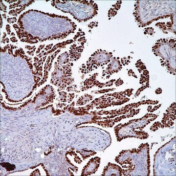
Cytokeratin 7 (OV-TL 12/30) Mouse Monoclonal Antibody

Sign Into View Organizational & Contract Pricing


About This Item

UNSPSC Code:
12352203
NACRES:
NA.41

biological source

mouse

Quality Level

100
500

conjugate

unconjugated

antibody form

diluted ascites fluid

antibody product type

primary antibodies

clone

OV-TL 12/30, monoclonal

description

For In Vitro Diagnostic Use in Select Regions (See Chart)

form

buffered aqueous solution

species reactivity

human

packaging

vial of 0.1 mL concentrate (307M-94)
vial of 0.5 mL concentrate (307M-95)
bottle of 1.0 mL predilute (307M-97)
vial of 1.0 mL concentrate (307M-96)
bottle of 7.0 mL predilute (307M-98)

manufacturer/tradename

Cell Marque

technique(s)

immunohistochemistry (formalin-fixed, paraffin-embedded sections): 1:100-1:500

isotype

IgG1κ

control

lung adenocarcinoma

shipped in

wet ice

storage temp.

2-8°C

visualization

cytoplasmic

Gene Information

human ... KRT7(3855)

General description

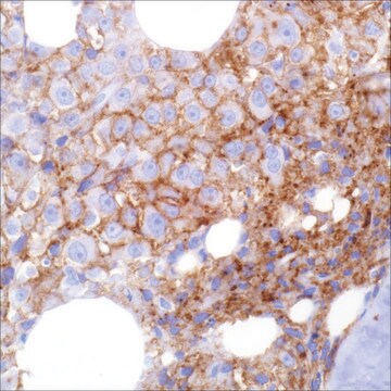
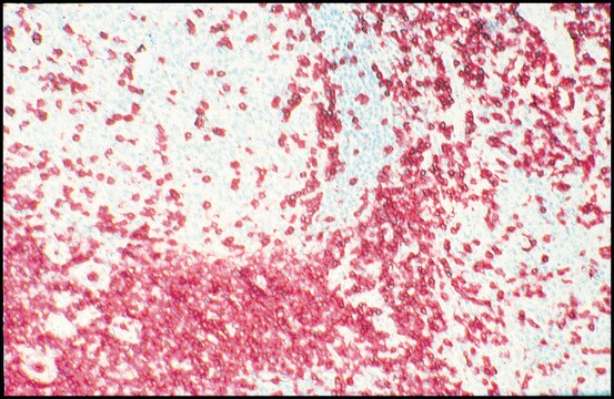
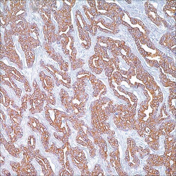
Anti-cytokeratin 7 reacts with proteins that are found in most ductal, glandular, transitional, and biliary duct epithelial cells. Cytokeratin 7 (CK7) labeling can help distinguish between lung, breast carcinomas, and urothelial carcinomas that are typically stain positive, and colon and prostate carcinomas that typically lack CK7 expression. CK7 is a common marker of primary lung adenocarcinomas and is observed in other primary lung carcinomas and non-pulmonary carcinomas. Anti-cytokeratin 7 has also been useful in the differential diagnosis of ovarianneoplasms.

Quality


IVD

IVD

IVD

RUO

Linkage

Cytokeratin 7 Positive Control Slides, Product No. 307S, are available for immunohistochemistry (formalin-fixed, paraffin-embedded sections).

Physical form

Solution in Tris Buffer, pH 7.3-7.7, with 1% BSA and <0.1% Sodium Azide

Preparation Note

Download the IFU specific to your product lot and formatNote: This requires a keycode which can be found on your packaging or product label.

Other Notes

For Technical Service please contact: 800-665-7284 or email: service@cellmarque.com

Legal Information

Cell Marque is a trademark of Merck KGaA, Darmstadt, Germany

Not finding the right product?  

Try our Product Selector Tool.


Certificates of Analysis (COA)

Search for Certificates of Analysis (COA) by entering the products Lot/Batch Number. Lot and Batch Numbers can be found on a product’s label following the words ‘Lot’ or ‘Batch’.

Already Own This Product?

Find documentation for the products that you have recently purchased in the Document Library.

Visit the Document Library

W G McCluggage et al.
Histopathology, 47(3), 231-247 (2005-08-24)
The ovary is a common site of metastatic tumour. In many cases of ovarian metastasis there is a known history of malignancy but in other cases the ovarian tumour is the first manifestation of disease. In this review metastatic colorectal
Shawn K Murray et al.
The American journal of surgical pathology, 28(9), 1154-1162 (2004-08-19)
We recently identified metastatic prostate carcinoma (PCA) within perirectal lymph nodes (PLNs) from 2 patients undergoing abdominoperineal resection (APR) for rectal adenocarcinoma (RA). As this phenomenon has not been addressed by any studies in the literature and because these positive
Somak Roy et al.
Archives of pathology & laboratory medicine, 135(12), 1601-1605 (2011-12-02)
Primary adenocarcinoma of urinary bladder is an uncommon neoplasm and is a source of diagnostic confusion with adenocarcinomas arising in adjacent organs, especially colon. These tumors show varied histologic picture and degree of differentiation. Clinical association with bladder exstrophy and
Sanjay Logani et al.
The American journal of surgical pathology, 27(11), 1434-1441 (2003-10-25)
Ovarian tumors containing cells with transitional cell morphology are recognized in the 1999 World Health Organization classification of ovarian tumors and include benign Brenner tumor, borderline and malignant Brenner tumor, and transitional cell carcinoma. Recent immunohistochemical investigations have reached conflicting
Kaitlin C Fogg et al.
Cancer letters, 458, 92-101 (2019-05-28)
High-grade serous ovarian cancer (HGSOC) metastasizes when tumor spheroids detach from the primary tumor and re-attach throughout the peritoneal cavity. Once the cancer cells have implanted in these new sites, the development of metastatic lesions is dependent on the disaggregation

Articles

Colorectal cancer is the third most common cancer in both men and women. An estimated 136,000 cases of colorectal cancer are expected to occur in 2016.

Our team of scientists has experience in all areas of research including Life Science, Material Science, Chemical Synthesis, Chromatography, Analytical and many others.

Contact Technical Service