Skip to Content
Merck
All Photos(1)

Key Documents

F8771

Sigma-Aldrich

Anti-Mouse IgG (Fab specific)−FITC antibody produced in goat

affinity isolated antibody, buffered aqueous solution

Synonym(s):

Goat Anti-Mouse IgG (Fab specific)−Fluorescein isothiocyanate

Sign Into View Organizational & Contract Pricing


About This Item

MDL number:
UNSPSC Code:
12352203
NACRES:
NA.46

biological source

goat

Quality Level

conjugate

FITC conjugate

antibody form

affinity isolated antibody

antibody product type

secondary antibodies

clone

polyclonal

form

buffered aqueous solution

storage condition

protect from light

technique(s)

immunohistochemistry (formalin-fixed, paraffin-embedded sections): 1:200
indirect immunofluorescence: 1:64

storage temp.

−20°C

target post-translational modification

unmodified

Looking for similar products? Visit Product Comparison Guide

General description

IgG antibody subtype is the most abundant serum immunoglobulins of the immune system. It is secreted by B cells and is found in blood and extracellular fluids and provides protection from infections caused by bacteria, fungi and viruses. Maternal IgG is transferred to fetus through the placenta that is vital for immune defence of the neonate against infections.

Specificity

Anti-Mouse IgG (Fab specific)-FITC antibody is specific for Fab fragment of all mouse IgG subclasses. It shows no reactivity with mouse IgG, Fc fragment, or human IgG. Goat anti-Mouse IgG is conjugated to FITC and then purified by gel filtration to remove free FITC. The antibody is adsorbed with human IgG and rat serum proteins to ensure minimal cross reactivity.

Immunogen

Purified mouse IgG

Application

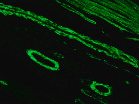
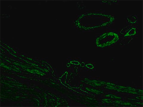
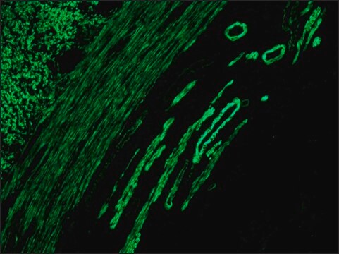
Anti-Mouse IgG (Fab specific)-FITC antibody may be used for immunofluorescence of human peripheral blood lymphocytes at a minimum dilution of 1:64. It may be used for immunohistochemistry of formalin-fixed, paraffin-embedded human tonsil slides at a working antibody dilution of 1:200. For immunofluorescence, a working dilution of 1:200 was used in various studies. To study DNA synthesis in mouse two-cell stage embryos by BrdU antibody, mouse IgG (Fab specific)-FITC antibody dilution of 1:50 was used.

Other Notes

Antibody adsorbed with human IgG and rat serum proteins.

Physical form

Solution in 0.01 M phosphate buffered saline, pH 7.4, containing 15 mM sodium azide.

Preparation Note

Adsorbed to reduce background with human or rat samples

Disclaimer

Unless otherwise stated in our catalog or other company documentation accompanying the product(s), our products are intended for research use only and are not to be used for any other purpose, which includes but is not limited to, unauthorized commercial uses, in vitro diagnostic uses, ex vivo or in vivo therapeutic uses or any type of consumption or application to humans or animals.

Not finding the right product?  

Try our Product Selector Tool.

Storage Class Code

10 - Combustible liquids

WGK

nwg

Flash Point(F)

Not applicable

Flash Point(C)

Not applicable

Personal Protective Equipment

dust mask type N95 (US), Eyeshields, Gloves

Choose from one of the most recent versions:

Certificates of Analysis (COA)

Lot/Batch Number

Don't see the Right Version?

If you require a particular version, you can look up a specific certificate by the Lot or Batch number.

Already Own This Product?

Find documentation for the products that you have recently purchased in the Document Library.

Visit the Document Library

María Florencia Sampedro et al.
F1000Research, 7, 1489-1489 (2018-11-27)
Background: E-cadherin is the major adhesion receptor in epithelial adherens junctions (AJs). On established epidermis, E-cadherin performs fine-tuned cell-cell contact remodeling to maintain tissue integrity, which is characterized by modulation of cell shape, size and packing density. In zebrafish, the
Sigrid Eckardt et al.
Reproduction (Cambridge, England), 129(5), 547-556 (2005-04-28)
Mammalian somatic cell cloning requires factors specific to the oocyte for reprogramming to succeed. This does not exclude that reprogramming continues during the zygote and cleavage stages. The capacity or role of zygotic and cleavage stages to reprogram somatic cell
P L Gabbott et al.
The Journal of comparative neurology, 350(2), 281-301 (1994-12-08)
The rationale for this study was to provide a comprehensive light microscopical description of the morphology of diaphorase-reactive neurons and neuropil elements in the dorsal lateral geniculate nucleus (dLGN) of the rat. An additional objective was to quantitatively assess whether
German Kamalov et al.
Journal of cardiovascular pharmacology, 62(6), 497-506 (2013-10-03)
Cardinal pathological features of hypertensive heart disease (HHD) include not only hypertrophied cardiomyocytes and foci of scattered microscopic scarring, a footprint of prior necrosis, but also small myocytes ensnared by fibrillar collagen where disuse atrophy with protein degradation would be
Elzbieta Pamula et al.
Journal of biomedical materials research. Part A, 89(2), 432-443 (2008-04-24)
Degradable three-dimensional porous scaffolds applicable as cell carriers for bone tissue engineering were developed by an innovative solvent casting/particulate leaching technique from poly(L-lactide-co-glycolide) (PLG). Three types of PLG scaffolds were prepared, and these had the same high porosity (83%) but

Our team of scientists has experience in all areas of research including Life Science, Material Science, Chemical Synthesis, Chromatography, Analytical and many others.

Contact Technical Service