Skip to Content
Merck
All Photos(1)

Key Documents

P9318

Sigma-Aldrich

Monoclonal Anti-Plectin antibody produced in mouse

clone 7A8, ascites fluid

Synonym(s):

Anti-EBS1, Anti-EBS5A, Anti-EBS5B, Anti-EBS5C, Anti-EBS5D, Anti-EBSMD, Anti-EBSND, Anti-EBSO, Anti-EBSOG, Anti-EBSPA, Anti-HD1, Anti-LGMD2Q, Anti-LGMDR17, Anti-PCN, Anti-PLEC1, Anti-PLEC1b, Anti-PLTN

Sign Into View Organizational & Contract Pricing


About This Item

MDL number:
UNSPSC Code:
12352203
NACRES:
NA.41

biological source

mouse

conjugate

unconjugated

antibody form

ascites fluid

antibody product type

primary antibodies

clone

7A8, monoclonal

mol wt

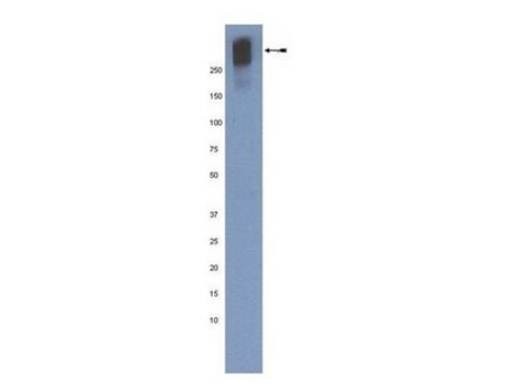
antigen 300 kDa

contains

15 mM sodium azide

species reactivity

marsupial (Ptoorous tridactylis PtK2 cell line), rat

technique(s)

dot blot: suitable
immunocytochemistry: suitable
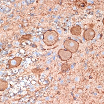
immunohistochemistry (frozen sections): suitable
indirect ELISA: suitable
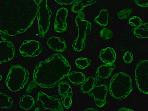
indirect immunofluorescence: 1:200 using unfixed frozen sections of rat heart
microarray: suitable
western blot: suitable

isotype

IgG1

shipped in

dry ice

storage temp.

−20°C

target post-translational modification

unmodified

Gene Information

General description

Monoclonal Anti-Plectin (mouse IgG1 isotype) is derived from the 7A8 hybridoma produced by the fusion of mouse myeloma cells and splenocytes from an immunized mouse. Plectin is an abundant, high molecular weight, cytomatrix protein (300 kDa) found in a wide variety of tissue and cell types. Plectin is found in stratified and non-stratified epithelia, fibroblasts, endothelial cells and astrocytes, as well as in striated, smooth and cardiac muscle, but not in neurons.

Specificity

The antibody recognizes an epitope located in the middle section of the rod domain of the plectin molecule, approximately halfway between the globular end domains. The kinase C phosphorylation site was found on the same terminal segment as this antibody epitope. The antibody specifically labels the 300 kDa band of plectin, however, lower M.W. band(s) may also be detected. The antibody stains filamentous structures on immunohistochemical staining of frozen tissue sections. The antibody inhibits the interaction of plectin with vimentin and lamin B. An additional application for this antibody is immunoelectronmicroscropy.

Immunogen

plectin from rat glioma C6 cells

Application

Monoclonal Anti-Plectin antibody produced in mouse has been used in:
  • ELISA
  • immunoblot
  • dot blot
  • immunocytochemistry
  • immunoelectron microscopy

Biochem/physiol Actions

Plectin plays a role in the cross-linking of intermediate filaments, the interlinking of intermediate filaments with microtubules and microfilaments and the anchoring of intermediate filaments to the plasma membrane and the nuclear membrane. It is a part of the signal transduction mechanism involving kinases A and C, because in vitro as well as in vivo phosphorylation of the protein by those kinases differentially affected its binding affinities to vimentin and lamin B.

Disclaimer

Unless otherwise stated in our catalog or other company documentation accompanying the product(s), our products are intended for research use only and are not to be used for any other purpose, which includes but is not limited to, unauthorized commercial uses, in vitro diagnostic uses, ex vivo or in vivo therapeutic uses or any type of consumption or application to humans or animals.

Not finding the right product?  

Try our Product Selector Tool.

Storage Class Code

10 - Combustible liquids

WGK

nwg

Flash Point(F)

Not applicable

Flash Point(C)

Not applicable


Certificates of Analysis (COA)

Search for Certificates of Analysis (COA) by entering the products Lot/Batch Number. Lot and Batch Numbers can be found on a product’s label following the words ‘Lot’ or ‘Batch’.

Already Own This Product?

Find documentation for the products that you have recently purchased in the Document Library.

Visit the Document Library

T M Svitkina et al.
The Journal of cell biology, 135(4), 991-1007 (1996-11-01)
By immunogold labeling, we demonstrate that "millipede-like" structures seen previously in mammalian cell cytoskeletons after removal of actin by treatment with gelsolin are composed of the cores of vimentin IFs with sidearms containing plectin. These plectin sidearms connect IFs to
Plectin-1 as a novel biomarker for pancreatic cancer
Bausch D, et al.
Clinical Cancer Research, 17(2), 302-309 (2011)
Plectin isoforms as organizers of intermediate filament cytoarchitecture
Wiche G and Winter L
Bioarchitecture, 1(1), 14-20 (2011)
Daisuke Tsuruta et al.
The Journal of biological chemistry, 278(40), 38707-38714 (2003-07-18)
Within each hemidesmosome, alpha6beta4 integrin plays a crucial role in hemidesmosome assembly by binding to laminin-5 in the basement membrane zone of epithelial tissue. Recent analyses have implicated "specificity-determining loops" (SDLs) in the I-like domain of beta integrin in regulating
Kiyoko Uehara et al.
Anatomical record (Hoboken, N.J. : 2007), 293(12), 2034-2043 (2010-11-23)
The ultrastructural distribution of vimentin intermediate filaments (IFs) and localizations of the related proteins in sinus endothelial cells of the rat spleen was examined by confocal laser scanning and electron microscopy with detergent extraction, myosin-fragment 1 decoration, and immunogold labeling

Our team of scientists has experience in all areas of research including Life Science, Material Science, Chemical Synthesis, Chromatography, Analytical and many others.

Contact Technical Service