Skip to Content
Merck
All Photos(2)

Key Documents

S2644

Sigma-Aldrich

Anti-S-100 antibody produced in rabbit

IgG fraction of antiserum, buffered aqueous solution

Synonym(s):

Anti-42C, Anti-ANX2LG, Anti-CAL1L, Anti-CLP11, Anti-Ca[1], Anti-GP11, Anti-P11, Anti-p10

Sign Into View Organizational & Contract Pricing


About This Item

MDL number:
UNSPSC Code:
12352203
NACRES:
NA.41

biological source

rabbit

conjugate

unconjugated

antibody form

IgG fraction of antiserum

antibody product type

primary antibodies

clone

polyclonal

form

buffered aqueous solution

species reactivity

rat, bovine, guinea pig, human

technique(s)

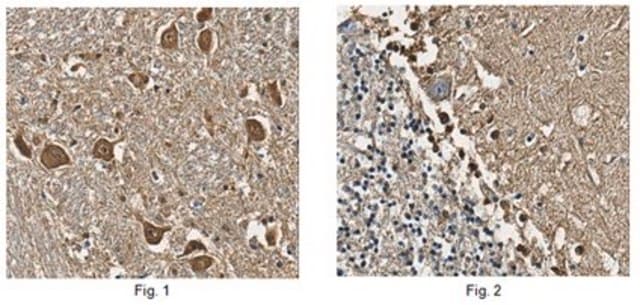
immunohistochemistry (formalin-fixed, paraffin-embedded sections): 20-100 μg/mL using sections of human or rat cerebellum
microarray: suitable

UniProt accession no.

shipped in

dry ice

storage temp.

−20°C

target post-translational modification

unmodified

Gene Information

General description

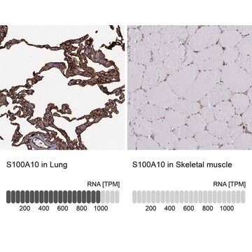
S-100 (S100 calcium binding protein A10) belongs to the EF-hand-containing S100 protein family. It is also termed as calpactin light chain, p11 or annexin 2 light chain. This gene is located on human chromosome 1q21.
S100 calcium binding protein A10 (S-100) is located on rat chromosome 2q34. It is also termed as p11.
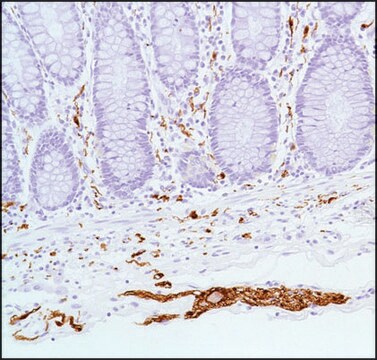
The S-100 protein family consists of around 16 calcium binding proteins that have distinct tissue specific expression patterns. S-100 proteins are widely expressed in neural and non-neural cell types, as well as in several cancers. These proteins regulate a broad range of cellular functions such as cell growth, signaling, cell structure, and energy metabolism . In immunohistology, the product specifically stains the S-100 protein. The product binds to S-100 protein in human, rat, guinea pig, and bovine. In the brain, the antibody can be used to stain glial and ependymal cells, as well as Schwann cells of the peripheral nervous system. In the skin, melanocytes and Langerhans cells can be stained by the antibody product. In lymph nodes, interdigitating reticulum cells are positively stained by anti-S-100 antibody. S-100 staining can be observed in almost all human benign and malignant melanocytic tumors.

Immunogen

S-100 from bovine brain

Application

Anti-S-100 antibody has been used in immunocytochemistry,[25} immunofluorescence and immunostaining.
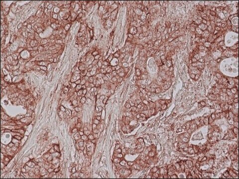
Anti-S-100 antibody is suitable for use in microarray, and immunohistochemistry (in formalin-fixed, paraffin-embedded sections, or using a conc. of 20-100 μg/mL in sections of human or rat cerebellum). The antibody may also be used in immunoelectrophoresis.

Biochem/physiol Actions

S-100 (S100 calcium binding protein A10) is required for trafficking of TRPV5 and TRPV6 (epithelial Ca2+ channels). S100A10 along with annexin 2, participates in several biological processes such as endocytosis, exocytosis and membrane-cytoskeleton interactions. It controls plasma membrane ion channels and cytosolic phospholipase A2.
S100 calcium binding protein A10 (S-100) plays an important role in depression-like character in rodents. In rat ventricular cardiomyocytes, p11 regulates calcium handling through 5-HT4R pathway.

Physical form

Solution in 0.01 M phosphate buffered saline, pH 7.4, containing 15 mM sodium azide.

Disclaimer

Unless otherwise stated in our catalog or other company documentation accompanying the product(s), our products are intended for research use only and are not to be used for any other purpose, which includes but is not limited to, unauthorized commercial uses, in vitro diagnostic uses, ex vivo or in vivo therapeutic uses or any type of consumption or application to humans or animals.

Not finding the right product?  

Try our Product Selector Tool.

Storage Class Code

10 - Combustible liquids

WGK

WGK 3

Flash Point(F)

Not applicable

Flash Point(C)

Not applicable


Certificates of Analysis (COA)

Search for Certificates of Analysis (COA) by entering the products Lot/Batch Number. Lot and Batch Numbers can be found on a product’s label following the words ‘Lot’ or ‘Batch’.

Already Own This Product?

Find documentation for the products that you have recently purchased in the Document Library.

Visit the Document Library

Identification of a peptide that interacts with Nestin protein expressed in brain cancer stem cells.
Beck S, et al.
Biomaterials, 32(33), 8518-8528 (2011)
Gene expression profile linked to p53 status in hepatitis C virus?related hepatocellular carcinoma.
Okada T, et al.
Febs Letters, 555(3), 583-590 (2003)
Cells of primarily extravalvular origin in degenerative aortic valves and bioprostheses
Skowasch D, et al.
European Heart Journal, 26 (2005)
Phospholipid-associated Annexin A2-S100A10 Heterotetramer and Its Subunits characterization of the interaction with tissue plasminogen activator, plasminogen, and plasmin.
MacLeod TJ, et al.
The Journal of Biological Chemistry, 278(28), 25577-25584 (2003)
Study of expression patterns and levels of neurotrophins and neurotrophin receptors in ulcerative colitis
Johansson M, et al.
Inflammatory bowel diseases, 13(4), 398-409 (2007)

Our team of scientists has experience in all areas of research including Life Science, Material Science, Chemical Synthesis, Chromatography, Analytical and many others.

Contact Technical Service