Skip to Content
Merck
All Photos(1)

Key Documents

05-224

Sigma-Aldrich

Anti-p53 Antibody, clone BP53-12

clone BP53-12, Upstate®, from mouse

Synonym(s):

Anti-BCC7, Anti-BMFS5, Anti-LFS1, Anti-P53, Anti-TRP53

Sign Into View Organizational & Contract Pricing


About This Item

UNSPSC Code:
12352203
eCl@ss:
32160702
NACRES:
NA.41

biological source

mouse

Quality Level

antibody form

purified immunoglobulin

antibody product type

primary antibodies

clone

BP53-12, monoclonal

species reactivity

human

packaging

antibody small pack of 25 μL

manufacturer/tradename

Upstate®

technique(s)

immunohistochemistry: suitable
immunoprecipitation (IP): suitable
western blot: suitable

isotype

IgG2a

NCBI accession no.

UniProt accession no.

shipped in

dry ice

target post-translational modification

unmodified

Gene Information

human ... TP53(7157)

General description

The human wild-type p53 protein is a 393 amino acid nuclear phosphoprotein, present in the nucleus of all normal mammalian cells where it appears to be involved in the regulation of cell proliferation. The normal protein has a very short half-life and is present in only minute amounts in normal tissues and cells. In contrast, mutant p53 protein produced by malignant cells is usually a product of a point mutation in the p53 gene leading to substitution of a single amino acid that significantly prolongs the half-life of the protein. The accumulation of high levels of p53 is a potential novel marker for malignancy.

Specificity

Recognizes p53.

Immunogen

Recombinant human wild type p53

Application

Anti-p53 Antibody, clone BP53-12 is a high quality Mouse Monoclonal Antibody for the detection of p53 & has been validated in IHC, IP & WB.
Research Category
Epigenetics & Nuclear Function
Research Sub Category
Transcription Factors

Quality

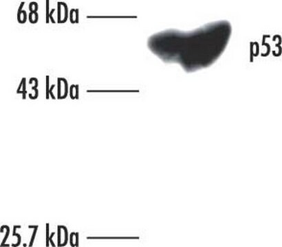
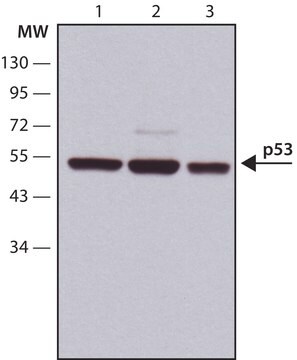
routinely evaluated on RIPA lysate of human Raji cells

Target description

53 kDa

Linkage

Replaces: 04-1083

Physical form

Format: Purified
Protein A purified
Protein G Purified immunoglobulin in Protein G Purified immunoglobulin in of 0.1M Tris-glycine, pH 7.4, 0.15M NaCl, 0.05% sodium azide before the addition of glycerol to 30%

Storage and Stability

Maintain for 2 years at -20°C from date of shipment. Aliquot to avoid repeated freezing and thawing. For maximum recovery of product, centrifuge the original vial after thawing and prior to removing the cap.

Analysis Note

Control
Raji cell lysate

Other Notes

Concentration: Please refer to the Certificate of Analysis for the lot-specific concentration.

Legal Information

UPSTATE is a registered trademark of Merck KGaA, Darmstadt, Germany

Disclaimer

Unless otherwise stated in our catalog or other company documentation accompanying the product(s), our products are intended for research use only and are not to be used for any other purpose, which includes but is not limited to, unauthorized commercial uses, in vitro diagnostic uses, ex vivo or in vivo therapeutic uses or any type of consumption or application to humans or animals.

Not finding the right product?  

Try our Product Selector Tool.

Storage Class Code

10 - Combustible liquids

WGK

WGK 1


Certificates of Analysis (COA)

Search for Certificates of Analysis (COA) by entering the products Lot/Batch Number. Lot and Batch Numbers can be found on a product’s label following the words ‘Lot’ or ‘Batch’.

Already Own This Product?

Find documentation for the products that you have recently purchased in the Document Library.

Visit the Document Library

Tiangui Chen et al.
Molecular medicine reports, 20(2), 1443-1450 (2019-06-08)
Autophagy is a conserved catabolic process by which cytoplasmic components are delivered into lysosomes for degradation. Pigment epithelium‑derived factor (PEDF) has been reported to be associated with autophagy and can induce p53 expression; however, the mechanism relating PEDF with autophagy
Translocation of p53 to mitochondria is regulated by its lipid binding property to anionic phospholipids and it participates in cell death control.
Ching-Hao Li,Yu-Wen Cheng,Po-Ling Liao,Jaw-Jou Kang
Neoplasia null
Dan-Dan Li et al.
PloS one, 7(9), e45058-e45058 (2012-10-02)
Topotecan produces DNA damage that induces autophagy in cancer cells. In this study, sensitising topotecan to colon cancer cells with different P53 status via modulation of autophagy was examined. The DNA damage induced by topotecan treatment resulted in cytoprotective autophagy
The estrogen receptor alpha pathway induces oncogenic Wip1 phosphatase gene expression.
Hye-Sook Han, Eunsil Yu, Ji-Young Song, Ji-Young Park, Se Jin Jang, Jene Choi et al.
Molecular Cancer Research null
Differential effect of hypoxia on etoposide-induced DNA damage response and p53 regulation in different cell types.
Sermeus, Audrey, et al.
Journal of Cellular Physiology (2013)

Our team of scientists has experience in all areas of research including Life Science, Material Science, Chemical Synthesis, Chromatography, Analytical and many others.

Contact Technical Service