16-204
Anti-Phosphotyrosine Antibody, recombinant clone 4G10®, biotin conjugate
clone 4G10®, Upstate®, from mouse
Sign Into View Organizational & Contract Pricing
All Photos(1)
About This Item
UNSPSC Code:
12352203
eCl@ss:
32160702
NACRES:
NA.41
conjugate:
biotin conjugate
application:
WB
clone:
4G10®, monoclonal
technique(s):
western blot: suitable
citations:
1
Recommended Products
biological source
mouse
Quality Level
conjugate
biotin conjugate
antibody form
purified antibody
antibody product type
primary antibodies
clone
4G10®, monoclonal
species reactivity (predicted by homology)
all
manufacturer/tradename
Upstate®
technique(s)
western blot: suitable
isotype
IgG2bκ
shipped in
wet ice
target post-translational modification
unmodified
Gene Information
human ... PID1(55022)
General description
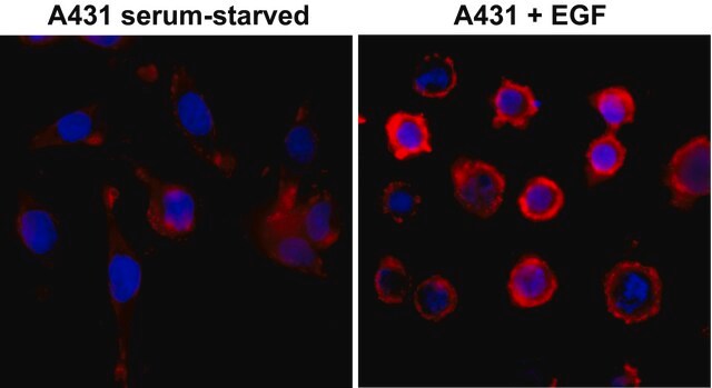
Included Positive Antigen Control: Catalog # 12-302, EGF-stimulated A431 cell lysate
Product Description: Produced from CHO cells expressing the 4G10 antibody heavy and light chain cDNAs. Heavy chain C-terminus has a hexa-histidine tag for purification and immobilization via nickel affinity matrices. Cross-linked to biotin. Patent pending.
Product Description: Produced from CHO cells expressing the 4G10 antibody heavy and light chain cDNAs. Heavy chain C-terminus has a hexa-histidine tag for purification and immobilization via nickel affinity matrices. Cross-linked to biotin. Patent pending.
Produced from CHO cells expressing the 4G10 antibody heavy and light chain cDNAs. Heavy chain C-terminus has a hexa-histidine tag for purification and immobilization via nickel affinity matrices. Cross-linked to biotin. Patent pending.
Specificity
Tyrosine-phosphorylated proteins from all species.
Immunogen
Phosphotyramine-KLH
Application
Detect Phosphotyrosine using this Anti-Phosphotyrosine Antibody, recombinant clone 4G10, biotin conjugate validated for use in WB.
Research Category
Signaling
Signaling
Research Sub Category
General Post-translation Modification
General Post-translation Modification
Quality
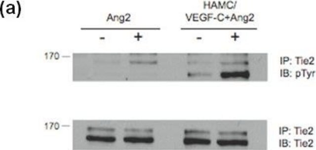
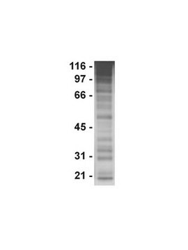
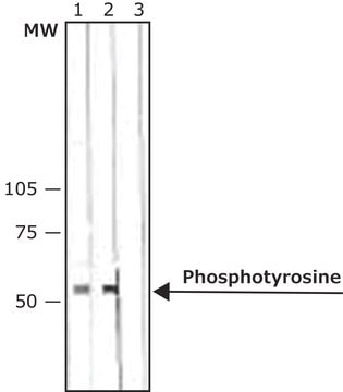
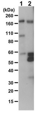
Routinely evaluated by western blot to detect tyrosine phosphorylated proteins in a RIPA lysate of EGF-stimulated A431 carcinoma cells.
Western Blot Analysis:
0.5-2 μg/mL of this lot detected tyrosine phosphorylated proteins in a RIPA lysate of EGF-stimulated A431 carcinoma cells.
Western Blot Analysis:
0.5-2 μg/mL of this lot detected tyrosine phosphorylated proteins in a RIPA lysate of EGF-stimulated A431 carcinoma cells.
Target description
Dependent upon the molecular weight of the tyrosine phosphorylated protein being detected.
Physical form
Biotin-conjugated recombinant mouse IgG2bκ in PBS containing 0.05% sodium azide. Liquid at 2-8°C.
Protein G Purified
Storage and Stability
Stable for 9 months at 2-8ºC from date of receipt.
For maximum recovery of product, centrifuge the vial prior to removing the cap. DO NOT FREEZE!
For maximum recovery of product, centrifuge the vial prior to removing the cap. DO NOT FREEZE!
Analysis Note
Control
Positive Antigen Control: Catalog #12-302, EGF-stimulated A431 cell lysate. Add 2.5µL of 2-mercaptoethanol/100µL of lysate and boil for 5 minutes to reduce the preparation. Load 20µg of reduced lysate per lane for minigels.
Positive Antigen Control: Catalog #12-302, EGF-stimulated A431 cell lysate. Add 2.5µL of 2-mercaptoethanol/100µL of lysate and boil for 5 minutes to reduce the preparation. Load 20µg of reduced lysate per lane for minigels.
Other Notes
Concentration: Please refer to the Certificate of Analysis for the lot-specific concentration.
Legal Information
4G10 is a registered trademark of Upstate Group, Inc.
UPSTATE is a registered trademark of Merck KGaA, Darmstadt, Germany
Disclaimer
Unless otherwise stated in our catalog or other company documentation accompanying the product(s), our products are intended for research use only and are not to be used for any other purpose, which includes but is not limited to, unauthorized commercial uses, in vitro diagnostic uses, ex vivo or in vivo therapeutic uses or any type of consumption or application to humans or animals.
Not finding the right product?
Try our Product Selector Tool.
Storage Class Code
10 - Combustible liquids
WGK
WGK 2
Certificates of Analysis (COA)
Search for Certificates of Analysis (COA) by entering the products Lot/Batch Number. Lot and Batch Numbers can be found on a product’s label following the words ‘Lot’ or ‘Batch’.
Already Own This Product?
Find documentation for the products that you have recently purchased in the Document Library.
Justin C Yarrow et al.
Chemistry & biology, 12(3), 385-395 (2005-03-31)
Small-molecule kinase inhibitors are predominantly discovered in pure protein assays. We have discovered an inhibitor of Rho-kinase (ROCK) through an image-based, high-throughput screen of cell monolayer wound healing. Using automated microscopy, we screened a library of approximately 16,000 compounds finding
Our team of scientists has experience in all areas of research including Life Science, Material Science, Chemical Synthesis, Chromatography, Analytical and many others.
Contact Technical Service
