Skip to Content
Merck
All Photos(3)

Key Documents

ABN1647

Sigma-Aldrich

Anti-Vesicular Glutamate Transporter 1 Antibody

rabbit polyclonal

Synonym(s):

Vesicular glutamate transporter 1, Brain-specific Na(+)-dependent inorganic phosphate cotransporter, Solute carrier family 17 member 7

Sign Into View Organizational & Contract Pricing


About This Item

UNSPSC Code:
12352203
eCl@ss:
32160702
NACRES:
NA.41

product name

Anti-VGluT1, from rabbit

biological source

rabbit

Quality Level

antibody form

unpurified

antibody product type

primary antibodies

clone

polyclonal

species reactivity

mouse, human, rat

species reactivity (predicted by homology)

bovine (based on 100% sequence homology)

packaging

antibody small pack of 25 μL

technique(s)

immunoautoradiography: suitable
immunofluorescence: suitable
immunohistochemistry: suitable
western blot: suitable

isotype

IgG

NCBI accession no.

UniProt accession no.

shipped in

ambient

target post-translational modification

unmodified

Gene Information

human ... SLC17A7(57030)

General description

Vesicular glutamate transporter 1 (UniProt: also known as VGluT1, Brain-specific Na(+)-dependent inorganic phosphate cotransporter, Solute carrier family 17 member 7) is encoded by the Slc17a7 (also known as Bnpi, Vglut1) gene (Gene ID: 116638) in rat. VGLUT1 is a multi-pass synaptic vesicle membrane protein that mediates the uptake of glutamate into synaptic vesicles at presynaptic nerve terminals of excitatory neuronal cells. Glutamate transporters mediate glutamate uptake into synaptic vesicles and are driven by a proton electrochemical gradient generated by the vacuolar H+-ATPase. Two isoforms of VGLUT1 have been reported that are produced by alternative splicing. VGLUT1 is expressed in the cerebellum, cerebral cortex and hippocampus. Expression of isoform VGLUT2 increases sharply during the first ten days of postnatal life, and also increases with increasing numbers of rhodopsin cells in the retina. It has been shown that both VGLUT1 and VGLUT2 are expressed prominently in the adult brain, however, during the first two weeks of postnatal development, VGLUT2 expression predominates. VGLUT1 expression becomes essential for survival at the time of the developmental switch from VGLUT2 to VGLUT1. The VGLUT1 knockout mice die at the age when a strong upregulation of VGLUT1 through newly emerging VGLUT1 synapses as well as a replacement of the previously predominant VGLUT2 isoform in cerebellar, hippocampal, and cortical neurons occurs. (Ref.: Wojcik, SM et al. (2004). Proc. Natl. Acad. Sci. USA 101(18): 7158-7163).

Specificity

This rabbit polyclonal antibody detects Vesicular glutamate transporter 1 in Human, Mouse, and Rat brain. It targets an epitope with in 14 amino acids from the C-terminal region.

Immunogen

Epitope: C-terminus
KLH-conjugated linear peptide corresponding to 14 amino acids from the C-terminal region of rat Vesicular glutamate transporter 1 (VGLUT1).

Application

Anti-VGLUT1, Cat. No. ABN1647, is a highly specific rabbit polyclonal antibody that targets Vesicular glutamate transporter 1 and has been tested in Immunoautoradiography, Immunofluorescence, Immunohistochemistry, and Western Blotting.
Immunoautoradiography Analysis: A representative lot detected VGLUT1 in rat and mouse striatum and hippocampus tissue (Courtesy of Dr.Salah el mestikawy PhD, at DR1 CNRS (CENTRE NATIONAL DE LA RECHERCHE SCIENTIFIQU - SATT LUTECH).

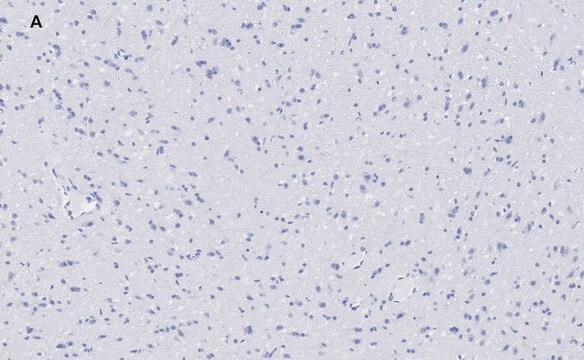
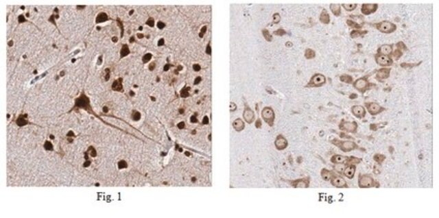
Immunohistochemistry Analysis: A representative lot detected VGLUT1 in prefrontal cortex of a representative control subject and AD patient (Kashani, A., et. al. (2008). Neurobiol Aging. 29(11):1619-30).

Western Blotting Analysis: A representative lot detected VGLUT1 in glutamate transporter 1 in rat and human putamen extracts (Kashani, A., et. al. (2007). Neurobiol Agining 28(4):568-78) and prefrontal cortex of controls, MCI, early stage Alzheimer (Kashani, A., et. al. (2008). Neurobiol Aging. 29(11):1619-30).

Immunofluorescence Analysis: A representative lot detected VGLUT1 in mouse striatum and hippocampus tissue (Courtesy of Dr.Salah el mestikawy PhD, at DR1 CNRS (CENTRE NATIONAL DE LA RECHERCHE SCIENTIFIQU - SATT LUTECH).

Immunofluorescence Analysis: A representative lot detected VGLUT1 in rat striatum (Herzog, E., et. al. (2001). J Neurosci. 21(22):RC181).

Immunoautoradiography Analysis: A representative lot detected VGLUT1 in coronal sections of frontal rat brain (Herzog, E., et. al. (2004). Neuroscience. 123(4):983-1002) and rat brain sagittal sections (Herzog, E., et. al. (2001). J Neurosci. 21(22):RC181).

Western Blotting Analysis: A representative lot detected VGLUT1 in BON cells stably expressing VGLUT1 (Herzog, E., et. al. (2001). J Neurosci. 21(22):RC181).
Research Category
Neuroscience

Quality

Evaluated by Western Blotting in mouse brain tissue lysate.

Western Blotting Analysis: A 1:1,000 dilution of this antibody detected VGLUT1 in 20 µg of mouse brain tissue lysate.

Target description

~55 kDa observed; 61.67 kDa calculated. Uncharacterized bands may be observed in some lysate(s).

Physical form

Format: Unpurified
Rabbit polyclonal antiserum with 0.05% sodium azide and 50% glycerol.
Unpurified

Storage and Stability

Stable for 1 year at -20°C from date of receipt. Handling Recommendations: Upon receipt and prior to removing the cap, centrifuge the vial and gently mix the solution. Aliquot into microcentrifuge tubes and store at -20°C. Avoid repeated freeze/thaw cycles, which may damage IgG and affect product performance.

Disclaimer

Unless otherwise stated in our catalog or other company documentation accompanying the product(s), our products are intended for research use only and are not to be used for any other purpose, which includes but is not limited to, unauthorized commercial uses, in vitro diagnostic uses, ex vivo or in vivo therapeutic uses or any type of consumption or application to humans or animals.

Not finding the right product?  

Try our Product Selector Tool.

Storage Class Code

12 - Non Combustible Liquids

WGK

WGK 1

Flash Point(F)

Not applicable

Flash Point(C)

Not applicable


Certificates of Analysis (COA)

Search for Certificates of Analysis (COA) by entering the products Lot/Batch Number. Lot and Batch Numbers can be found on a product’s label following the words ‘Lot’ or ‘Batch’.

Already Own This Product?

Find documentation for the products that you have recently purchased in the Document Library.

Visit the Document Library

Customers Also Viewed

Liqiang Chen et al.
eLife, 11 (2022-07-02)
The presynaptic protein α-synuclein (αSyn) has been suggested to be involved in the pathogenesis of Parkinson's disease (PD). In PD, the amygdala is prone to develop insoluble αSyn aggregates, and it has been suggested that circuit dysfunction involving the amygdala
Ken Matsuura et al.
iScience, 25(8), 104692-104692 (2022-07-21)
The CaMKΙΙα-Cre mouse lines, possibly the most used Cre lines in neuroscience, have resulted in over 800 articles to date. Here, we demonstrate that the second most widely used CaMKΙΙα-Cre line, Tg(Camk2a-cre)2Gsc (or CamiCre), shows ectopic overexpression of synaptotagmin 2
Hang Zhou et al.
British journal of anaesthesia, 126(6), 1141-1156 (2021-03-02)
Both animal and retrospective human studies have linked extended and repeated general anaesthesia during early development with cognitive and behavioural deficits later in life. However, the neuronal circuit mechanisms underlying this anaesthesia-induced behavioural impairment are poorly understood. Neonatal mice were
Yuzuha Shiraki et al.
Molecular biology of the cell, 34(11), ar110-ar110 (2023-08-16)
Alcadein α (Alcα) and amyloid-β protein precursor (APP) are cargo receptors that associate vesicles with kinesin-1. These vesicles, which contain either Alcα or APP, transport various proteins/cargo molecules into axon nerve terminals. Here, we analyzed immune-isolated Alcα- and APP-containing vesicles
Swagata Ghatak et al.
eLife, 8 (2019-11-30)
Human Alzheimer's disease (AD) brains and transgenic AD mouse models manifest hyperexcitability. This aberrant electrical activity is caused by synaptic dysfunction that represents the major pathophysiological correlate of cognitive decline. However, the underlying mechanism for this excessive excitability remains incompletely

Our team of scientists has experience in all areas of research including Life Science, Material Science, Chemical Synthesis, Chromatography, Analytical and many others.

Contact Technical Service