Skip to Content
Merck
All Photos(1)

Key Documents

268R-1

Sigma-Aldrich

IgD (EP173) Rabbit Monoclonal Primary Antibody

Sign Into View Organizational & Contract Pricing


About This Item

UNSPSC Code:
12352203
NACRES:
NA.41

biological source

rabbit

Quality Level

100
500

conjugate

unconjugated

antibody form

culture supernatant

antibody product type

primary antibodies

clone

EP173, monoclonal

description

(For In Vitro Diagnostic Use in Select Regions (See Chart))

form

buffered aqueous solution

species reactivity

human

packaging

bottle of 1.0 mL predilute (268R-17)
bottle of 7.0 mL predilute (268R-18)
vial of 0.1 mL concentrate (268R-14)
vial of 0.5 mL concentrate (268R-15)
vial of 1.0 mL concentrate (268R-16)

manufacturer/tradename

Cell Marque

technique(s)

immunohistochemistry (formalin-fixed, paraffin-embedded sections): 1:25-1:100

isotype

IgG

shipped in

wet ice

storage temp.

2-8°C

General description

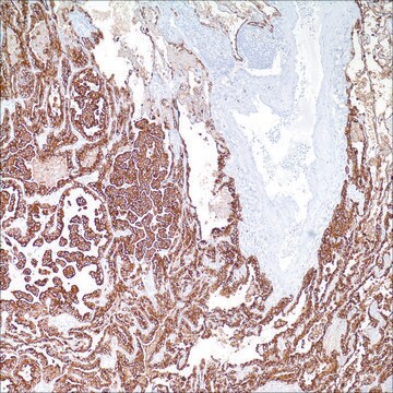
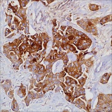
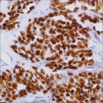
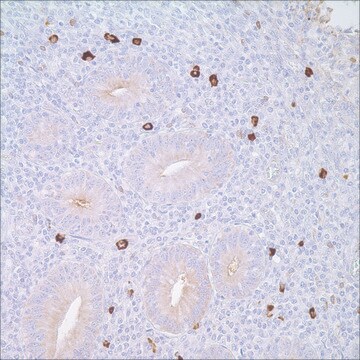
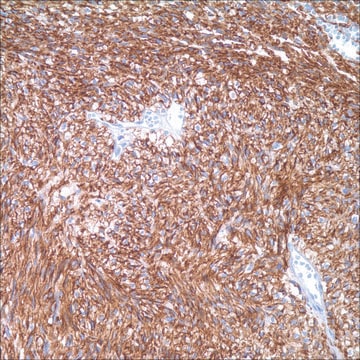
The monoclonal antibody against IgD reacts with immunoglobin D delta chains. In tonsil and lymph node, immunohistochemical staining for IgD immunoglobulin heavy chain is usually used to highlight the tonsil and nodal architecture since the IgD antibody stains mantle zone cells in secondary follicles and mantle cells in primary follicles.1 It has been reported that IgD can be detected in the surface/cytoplasm of neoplastic cells of common small B lymphoid cell lymphomas, such as small lymphocytic lymphoma, mantle cell lymphoma, marginal zone lymphoma (especially splenic marginal zone lymphoma), and follicular lymphoma.1-2 IgD expression in L & P cells of nodular lymphocyte predominant Hodkin lymphoma has been seen in subsets of cases (27% to 71.4%).1-3 The IgD positive L & P cells are usually located in the extrafollicular area with a relatively T-cell-rich background1,3 IgD expression is rarely seen in T-cell rich B-cell lymphoma. Studies have demonstrated that Reed-Sternberg cells of classic Hodgkin lymphoma were negative for IgD1,3 IgD multiple myeloma is a rare bone marrow plasma cell dyscrasia and can be identified by the IgD antibody, especially when a dry tap is encountered.4

Quality


IVD

IVD

IVD

RUO

Linkage

IgD Positive Control Slides, Product No. 268S, are available for immunohistochemistry (formalin-fixed, paraffin-embedded sections).

Physical form

Solution in Tris Buffer, pH 7.3-7.7, with 1% BSA and <0.1% Sodium Azide.

Preparation Note

Download the IFU specific to your product lot and formatNote: This requires a keycode which can be found on your packaging or product label.

Other Notes

For Technical Service please contact: 800-665-7284 or email: service@cellmarque.com

Legal Information

Cell Marque is a trademark of Merck KGaA, Darmstadt, Germany

Not finding the right product?  

Try our Product Selector Tool.

Storage Class Code

12 - Non Combustible Liquids

WGK

WGK 2

Flash Point(F)

Not applicable

Flash Point(C)

Not applicable


Choose from one of the most recent versions:

Certificates of Analysis (COA)

Lot/Batch Number

Don't see the Right Version?

If you require a particular version, you can look up a specific certificate by the Lot or Batch number.

Already Own This Product?

Find documentation for the products that you have recently purchased in the Document Library.

Visit the Document Library

Philip M Kluin et al.
The Journal of pathology, 236(3), 302-314 (2015-02-28)
Many hyperplasias and lymphomas of marginal zone B-cells are associated with infection. We identified six children and one adolescent with cervical lymphadenopathy showing prominent polyclonal nodal marginal zone hyperplasia (pNMZH) and four adolescents with monoclonal paediatric nodal marginal zone lymphoma
Shivlal Pandey et al.
Oncology (Williston Park, N.Y.), 27(8), 798-803 (2013-10-19)
Immunoglobulin D multiple myeloma (IgD MM) accounts for almost 2% of all myeloma cases. It is associated with an increased frequency of undetectable or small monoclonal (M)-protein levels on electrophoresis; osteolytic lesions; extramedullary involvement; amyloidosis; a lambda (lambda) light chain
Sonam Prakash et al.
The American journal of surgical pathology, 30(5), 585-592 (2006-05-16)
Nodular lymphocyte predominant Hodgkin lymphoma (NLPHL) is a rare B-cell lymphoma considered to be of germinal center (GC) derivation. Studies on immunoglobulin expression have been few, and post-switch immunoglobulin (IgG) has been identified in the majority of cases examined thus
Aliyah R Sohani et al.
The American journal of surgical pathology, 35(11), 1666-1678 (2011-10-15)
Nodular lymphocyte-predominant Hodgkin lymphoma (NLPHL) is a distinct Hodgkin lymphoma subtype composed of few neoplastic lymphocyte-predominant (LP) cells in a background of reactive small B and T cells. We have seen occasional NLPHL cases that contain background T cells with

Our team of scientists has experience in all areas of research including Life Science, Material Science, Chemical Synthesis, Chromatography, Analytical and many others.

Contact Technical Service