Skip to Content
Merck
All Photos(2)

Key Documents

F0397

Sigma-Aldrich

Anti-Cytokeratin pan-FITC antibody, Mouse monoclonal

clone PCK-26, purified from hybridoma cell culture

Synonym(s):

Anti-CK16-Fluorescein isothiocyanate, Anti-FNEPPK, Anti-K16, Anti-K1CP, Anti-KRT16A, Anti-NEPPK, Anti-PC1

Sign Into View Organizational & Contract Pricing


About This Item

UNSPSC Code:
12352203
NACRES:
NA.41

biological source

mouse

Quality Level

conjugate

FITC conjugate

antibody form

purified from hybridoma cell culture

antibody product type

primary antibodies

clone

PCK-26, monoclonal

form

buffered aqueous solution

technique(s)

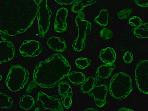
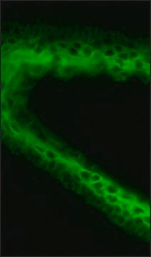
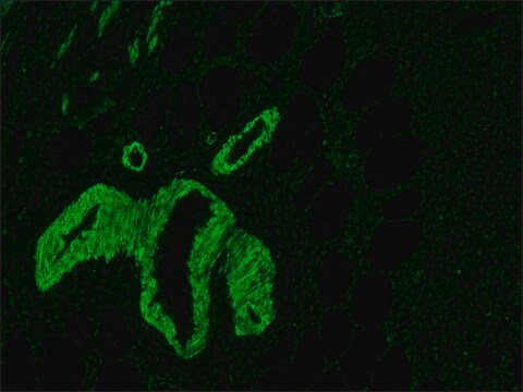
direct immunofluorescence: 1:25 using formalin-fixed, paraffin-embedded sections of human or animal tissues
immunohistochemistry (frozen sections): suitable

shipped in

dry ice

storage temp.

−20°C

target post-translational modification

unmodified

General description

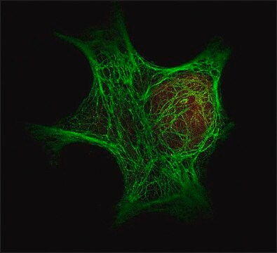
Cytokeratins are proteins of keratin-containing intermediate filaments that provide mechanical support and other additional functions in epithelial cells. It is found in the intracytoplasmic cytoskeleton of epithelial tissue. Epithelial tissue expresses cytokeratin subunits in a specific and stable pattern. Cytokeratins along with vimentin are involved in cell proliferation, migration and differentiation of preodontoblasts and preameloblasts. The intermediate-sized filaments are abundant in human endothelial cells and are mostly of vimentin type.
FITC Monoclonal Anti-Pan Cytokeratin is a purified mouse monoclonal antibody conjugated with fluorescein isothiocyanate (FITC) isomer I. Monoclonal anti-pan cytokeratin (mouse IgG1 isotype) is derived from the PCK-26 hybridoma produced by the fusion of mouse myeloma cells and splenocytes of immunized BALB/c mice. Cytokeratins, a group comprised of at least 29 different proteins are characteristic of epithelial and trichocytic cells. Cytokeratin 5, 6 and 8 are members of the type II, neutral-to-basic subfamily. Cytokeratin peptide 5 is the primary type II keratin in stratified epithelia while cytokeratin type 8 is a major type II keratin in simple epithelia. Cytokeratin 6 is a "hyperproliferation" cytokeratin expressed in tissues with natural or pathological high turnover.

Specificity

The antibody recognizes an epitope located on the Type II cytokeratins 1, 5, 6, and 8. PCK-26 is a broad spectrum antibody which reacts specifically with a variety of normal, reactive, and neoplastic epithelial tissues. The antibody reacts with simple, cornifying, and non-cornifying squamous epithelia and pseudostratified epithelia.

Immunogen

cytokeratin from human epidermis.

Application

Monoclonal Anti-Cytokeratin, pan-FITC antibody produced in mouse has been used in:
  • immunohistochemistry
  • staining chicken intestinal cells
  • immunofluorescence staining

Monoclonal Anti-Cytokeratin, pan-FITC antibody produced in mouse is suitable for direct immunofluorescence at a dilution of 1:25 using formalin-fixed, paraffin-embedded sections of human or animal tissues and immunohistochemistry (frozen sections).

Physical form

Solution in 0.01 M phosphate buffered saline, pH 7.4, containing 1% BSA and 15 mM sodium azide.

Disclaimer

Unless otherwise stated in our catalog or other company documentation accompanying the product(s), our products are intended for research use only and are not to be used for any other purpose, which includes but is not limited to, unauthorized commercial uses, in vitro diagnostic uses, ex vivo or in vivo therapeutic uses or any type of consumption or application to humans or animals.

Not finding the right product?  

Try our Product Selector Tool.

Storage Class Code

10 - Combustible liquids

WGK

nwg

Flash Point(F)

Not applicable

Flash Point(C)

Not applicable

Personal Protective Equipment

dust mask type N95 (US), Eyeshields, Gloves

Choose from one of the most recent versions:

Certificates of Analysis (COA)

Lot/Batch Number

Don't see the Right Version?

If you require a particular version, you can look up a specific certificate by the Lot or Batch number.

Already Own This Product?

Find documentation for the products that you have recently purchased in the Document Library.

Visit the Document Library

Miranda E Sowder et al.
Scientific reports, 8(1), 14299-14299 (2018-09-27)
Breast cancer cells frequently home to the bone, but the mechanisms controlling tumor colonization of the bone marrow remain unclear. We report significant enrichment of bone-disseminated estrogen receptor positive human MCF7 cells by 17 β-estradiol (E2) following intracardiac inoculation. Using flow
Takashi Maeda et al.
Cancer research, 64(2), 612-621 (2004-01-28)
Infiltrating carcinomas characteristically elicit a reactive stromal response, and accumulating evidence indicates that tumor stroma fibroblasts reciprocally promote tumor development and growth. The cell surface heparan sulfate proteoglycan, syndecan-1 (Sdc1), is thought to function as a coreceptor for growth factor
Interleukin 22 prevents lipopolysaccharide-induced preterm labor in mice
Dambaeva S, et al.
Biology of Reproduction, 98(3), 299-308 (2018)
Umida Ganieva et al.
Frontiers in immunology, 13, 955576-955576 (2022-09-13)
The uterine endometrium uniquely regenerates after menses, postpartum, or after breaks in the uterine layer integrity throughout women's lives. Direct cell-cell contacts ensured by tight and adherens junctions play an important role in endometrial integrity. Any changes in these junctions
Serial culture of murine primary airway epithelial cells and ex vivo replication of human rhinoviruses
Brockman-SRA, et al.
Journal of Immunological Methods, 339(2), 264-269 (2008)

Our team of scientists has experience in all areas of research including Life Science, Material Science, Chemical Synthesis, Chromatography, Analytical and many others.

Contact Technical Service