Skip to Content
Merck
All Photos(5)

Key Documents

F0916

Sigma-Aldrich

Anti-Fibronectin Antibody

mouse monoclonal, IST-4

Synonym(s):

Anti-CIG, Anti-ED-B, Anti-FINC, Anti-FN, Anti-FNZ, Anti-GFND, Anti-GFND2, Anti-LETS, Anti-MSF, Anti-SMDCF

Sign Into View Organizational & Contract Pricing


About This Item

MDL number:
UNSPSC Code:
12352203
NACRES:
NA.41

Product Name

Monoclonal Anti-Fibronectin antibody produced in mouse, clone IST-4, ascites fluid

biological source

mouse

Quality Level

conjugate

unconjugated

antibody form

ascites fluid

antibody product type

primary antibodies

clone

IST-4, monoclonal

mol wt

antigen 240 kDa

contains

15 mM sodium azide

species reactivity

human

should not react with

rabbit, mouse, goat, bovine, canine, chicken

technique(s)

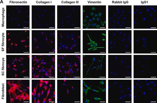
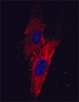
immunocytochemistry: 1:100 using cultured human fibroblasts
immunohistochemistry (formalin-fixed, paraffin-embedded sections): suitable
immunohistochemistry (frozen sections): suitable
indirect ELISA: suitable
radioimmunoassay: suitable
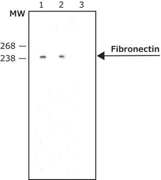
western blot: 1:250 using human fibroblasts cell extract

isotype

IgG1

UniProt accession no.

shipped in

dry ice

storage temp.

−20°C

target post-translational modification

unmodified

Gene Information

human ... FN1(2335)

Looking for similar products? Visit Product Comparison Guide

Related Categories

General description

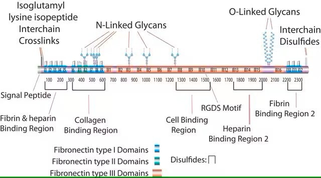
Fibronectin (FN) is an extracellular matrix protein composed of two nearly-identical disulfide-bound polypeptides with typical molecular weights of 220-280 kDa. Cellular fibronectin is structurally and antigenically similar to cold insoluble globulin from plasma and antibodies to either form usually cross-react. Careful analysis of the FN molecule indicates that it contains several functionally and structurally distinct domains which may bind to cell surfaces and to a variety of molecules such as collagen, heparin, gelatin, fibrin and DNA. FN′s play an important role in diverse biological phenomena including cell adhesion, cell migration, hemostasis and thrombosis, wound healing and the ability to induce a more normal phenotype in transformed cells.
Fibronectin 1 is a glycoprotein of the extracellular matrix that is coded by FN1 gene. It is expressed in the plasma and at the cell surface. It is mapped to human chromosome 2q35.
Mouse monoclonal clone IST-4 anti-Human Fibronectin antibody recognizes an epitope located within the 5th type III repeat of human plasma fibronectin. This epitope is common to all fibronectin forms. The antibody localizes plasma and cellular fibronectin both in its native and denatured-reduced forms (240 kDa) applying immunoblotting, RIA, ELISA, immunocytochemistry and immunohistochemistry. The product reacts with human fibronectin. No reactivity is observed with other species including goat, bovine, dog, rabbit, mouse and chicken.

Immunogen

human plasma fibronectin

Application

Applications in which this antibody has been used successfully, and the associated peer-reviewed papers, are given below.
Immunofluorescence (1 paper)
Immunohistochemistry (1 paper)
Western Blotting (1 paper)
Monoclonal Anti-Fibronectin antibody has been used :
  • in immunohistochemistry
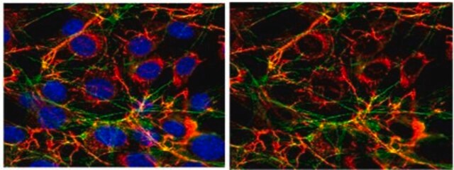
  • in confocal microscopy
  • in immunocytochemistry
  • western blotting analysis

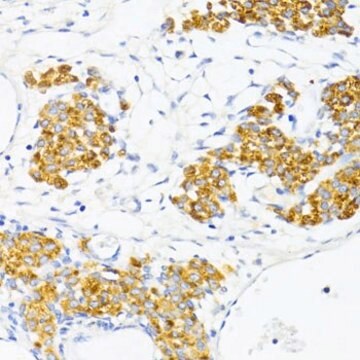
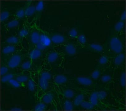
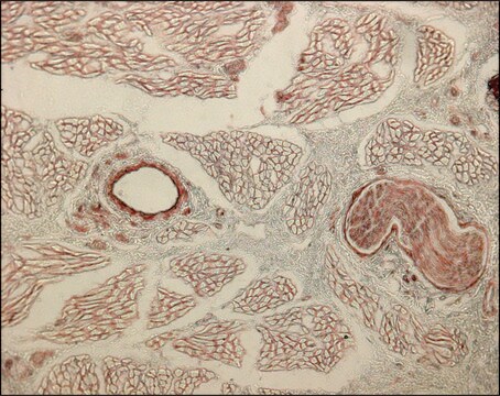
Mouse monoclonal clone IST-4 anti-Human Fibronectin antibody may be used for the specific labeling of fibronectin in cultured cells and in frozen tissue sections. Staining of formalin-fixed, paraffin-embedded sections may require prolonged proteolytic unmasking (e.g., by pepsin but not by trypsin).

Biochem/physiol Actions

Fibronectin participates in growth, blood coagulation and metastasis. Mutations in FN1 results in glomerulopathy. It plays an important role in cell attachment and spreading, control of cell cytoskeleton, morphology and differentiation. FN1 is also involved in the formation of extracellular matrix.

Disclaimer

Unless otherwise stated in our catalog or other company documentation accompanying the product(s), our products are intended for research use only and are not to be used for any other purpose, which includes but is not limited to, unauthorized commercial uses, in vitro diagnostic uses, ex vivo or in vivo therapeutic uses or any type of consumption or application to humans or animals.

Not finding the right product?  

Try our Product Selector Tool.

Storage Class Code

10 - Combustible liquids

WGK

nwg

Flash Point(F)

Not applicable

Flash Point(C)

Not applicable


Choose from one of the most recent versions:

Certificates of Analysis (COA)

Lot/Batch Number

Don't see the Right Version?

If you require a particular version, you can look up a specific certificate by the Lot or Batch number.

Already Own This Product?

Find documentation for the products that you have recently purchased in the Document Library.

Visit the Document Library

TAZ promotes cell proliferation and epithelial-mesenchymal transition and is inhibited by the hippo pathway
Lei Q Y, et al.
Molecular and cellular biology, 28(7), 2426-2436 (2008)
Francesca Di Modugno et al.
Oncogene, 37(42), 5605-5617 (2018-06-17)
We demonstrated previously that the splicing of the actin regulator, hMENA, generates two alternatively expressed isoforms, hMENA11a and hMENAΔv6, which have opposite functions in cell invasiveness. Their mechanisms of action have remained unclear. Here we report two major findings: (i)
The formation of a functional retinal pigment epithelium occurs on porous polytetrafluoroethylene substrates independently of the surface chemistry
Kearns V R, et al.
Journal of Materials Science. Materials in Medicine, 28(8), 124-124 (2017)
Ryan J Nagao et al.
Journal of materials chemistry. B, 1(37), 4801-4808 (2013-10-02)
Promoting regeneration using scaffolds created by decellularizing native tissue is becoming a popular technique applied to a variety of tissues. We demonstrate a method to decellularize highly vascular tissue keeping the vascular structure intact down to the capillary scale. Using
Clinicopathology of graft detachment after D escemet's membrane endothelial keratoplasty
Brockmann T, et al.
Acta Ophthalmologica, 92(7), e556-e561 (2014)

Protocols

TrueGel3D® Hydrogel Plate protocol guides high-throughput culture of human adipose MSCs for screening applications.

TrueGel3D® Hydrogel Plate protocol guides high-throughput culture of human adipose MSCs for screening applications.

TrueGel3D® Hydrogel Plate protocol guides high-throughput culture of human adipose MSCs for screening applications.

TrueGel3D® Hydrogel Plate protocol guides high-throughput culture of human adipose MSCs for screening applications.

Our team of scientists has experience in all areas of research including Life Science, Material Science, Chemical Synthesis, Chromatography, Analytical and many others.

Contact Technical Service