Skip to Content
Merck
All Photos(1)

Key Documents

M5293

Sigma-Aldrich

Monoclonal Anti-Mucin Gastric antibody produced in mouse

clone 45M1, ascites fluid

Sign Into View Organizational & Contract Pricing


About This Item

MDL number:
UNSPSC Code:
12352203
NACRES:
NA.41

biological source

mouse

Quality Level

conjugate

unconjugated

antibody form

ascites fluid

antibody product type

primary antibodies

clone

45M1, monoclonal

contains

15 mM sodium azide

species reactivity

chicken, monkey, rat, rabbit, pig, hedgehog, mouse, human, feline

technique(s)

immunocytochemistry: suitable
immunohistochemistry (formalin-fixed, paraffin-embedded sections): 1:200 using sections of human stomach
western blot: suitable using non-reducing conditions

isotype

IgG1

UniProt accession no.

shipped in

dry ice

storage temp.

−20°C

target post-translational modification

unmodified

Gene Information

human ... MUC5AC(4586)
mouse ... Muc5ac(17833)
rat ... Muc5ac(682837)

Related Categories

General description

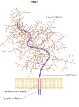
Monoclonal Anti-Human Gastric Mucin (mouse IgG1 isotype) is derived from the 445M1 hybridoma produced by the fusion of mouse myeloma cells and splenocytes from BALB/c mice immunized with mucin isolated from human ovarian cyst fluid. Mucin is a high M.W. (1,000 kDa) glycoprotein, expressed by mucus cells of the gastric epithelium and by goblet cells of the fetal, precancerous and cancerous colon, but not by those of the normal colon. It also appears in other epithelial tissues, which are embryologically derived from the foregut (epigastric and bronchial epithelium) and in Müller ducts (mucus cells of the endocervix and urethral epithelium near the prostatic utriculus).
Mucin has a netlike, branched structure and is a secreted protein. It belongs to the category of gel-forming mucins. The gene encoding this protein is upregulated by interleukin-1β (IL-1β) and is localized on chromosome 11p15.5.

Specificity

The antibody recognizes the mucin epitope g located in the peptide core of gastric mucin (>1,000 kDa). This epitope is completely destroyed by thiol reduction (using mercaptoethanol) and partially lost following trypsin proteolysis, but is stable upon periodate oxidation. The antibody stains the surface gastric epithelium of normal human gastrointestinal tract and reacts with fetal, pre-cancerous and cancerous colonic mucosa, but not with normal colon.

Immunogen

mucin from human ovarian cyst fluid.

Application

Applications in which this antibody has been used successfully, and the associated peer-reviewed papers, are given below.
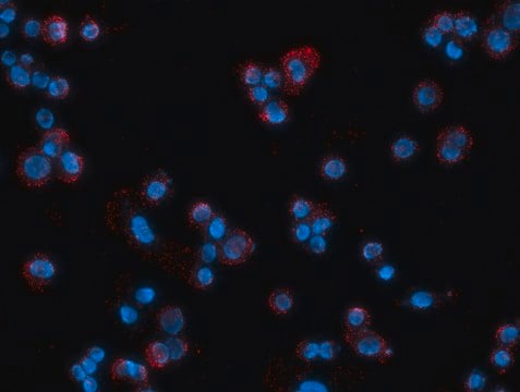
Immunofluorescence (1 paper)
Monoclonal Anti-Human Gastric Mucin may be used for the localization of gastric mucin using various immunochemical assays such as immunoblot, immunofluorescence, immunohistochemistry, immunostaining and immunoradiofixation.

Biochem/physiol Actions

Mucin is composed of a peptide core containing heavily glycosylated regions and nonglycosylated regions. The heavily glycosylated components of mucin consist of a mosaic of epitopes, some of which are associated with saccharide moieties and are related to blood-group antigens, and others which are M1 antigens associated with the peptidic core. M1 antigens are common to human ovarian mucinous cyst fluids and to gastric mucosa. M1 is distributed in various human tumors such as the colon, stomach, pancreas, ovary, Barrets′ esophagus, endocervix, and endometrium carcinomas.

Disclaimer

Unless otherwise stated in our catalog or other company documentation accompanying the product(s), our products are intended for research use only and are not to be used for any other purpose, which includes but is not limited to, unauthorized commercial uses, in vitro diagnostic uses, ex vivo or in vivo therapeutic uses or any type of consumption or application to humans or animals.

Not finding the right product?  

Try our Product Selector Tool.

Storage Class Code

10 - Combustible liquids

WGK

nwg

Flash Point(F)

Not applicable

Flash Point(C)

Not applicable


Certificates of Analysis (COA)

Search for Certificates of Analysis (COA) by entering the products Lot/Batch Number. Lot and Batch Numbers can be found on a product’s label following the words ‘Lot’ or ‘Batch’.

Already Own This Product?

Find documentation for the products that you have recently purchased in the Document Library.

Visit the Document Library

Emma C Skoog et al.
PloS one, 7(5), e36378-e36378 (2012-05-09)
Helicobacter pylori colonizes the mucus niche of the gastric mucosa and is a risk factor for gastritis, ulcers and cancer. The main components of the mucus layer are heavily glycosylated mucins, to which H. pylori can adhere. Mucin glycosylation differs
Optimized Protocol for Regeneration of the Conjunctival Epithelium Using the Cell Suspension Technique.
Bertolin M, et al.
Cornea, 38(4), 469-479 (2019)
Marina Bertolin et al.
Cornea, 38(4), 469-479 (2018-06-21)
To develop autologous tissue-engineered conjunctival epithelial sheets to be used as advanced therapy medicinal products for severe ocular surface disorders involving the conjunctiva. Methods used aimed at 1) mapping the conjunctiva for identification of the stem cell location, 2) establishing
MUC5AC upstream complex repetitive region length polymorphisms are associated with susceptibility and clinical stage of gastric cancer.
Wang C
PLoS ONE, 9(6), e98327-e98327 (2014)
The gastrointestinal mucus system in health and disease.
Johansson MEV, et al.
Nature Reviews. Gastroenterology & Hepatology, 10(6), 352-352 (2013)

Our team of scientists has experience in all areas of research including Life Science, Material Science, Chemical Synthesis, Chromatography, Analytical and many others.

Contact Technical Service