SAB4700040
Monoclonal Anti-CD3 antibody produced in mouse
clone OKT3, purified immunoglobulin, buffered aqueous solution
Synonym(s):
Mouse Anti-T-cell surface antigen T3/Leu-4 epsilon chain, Mouse Monoclonal Anti-T3E
Sign Into View Organizational & Contract Pricing
All Photos(1)
About This Item
UNSPSC Code:
12352203
NACRES:
NA.41
Recommended Products
biological source
mouse
Quality Level
conjugate
unconjugated
antibody form
purified immunoglobulin
antibody product type
primary antibodies
clone
OKT3, monoclonal
form
buffered aqueous solution
species reactivity
human
concentration
1 mg/mL
technique(s)
flow cytometry: suitable
isotype
IgG2a
NCBI accession no.
shipped in
wet ice
storage temp.
2-8°C
target post-translational modification
unmodified
Gene Information
human ... CD3(916)
General description
The CD3 gene is mapped to human chromosome 11q23.3. The encoded protein exists in three isoforms, CD3ε, CD3γ and CD3δ and each contains an N-terminal extracellular domain, a transmembrane segment and a cytoplasmic domain. CD3 is a 20kDa glycoprotein expressed on surface of all human T lymphocytes.
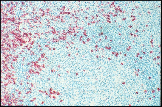
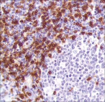
The mouse monoclonal antibody OKT3 recognizes the CD3 antigen of the TCR/CD3 complex on mature human T cells. This antibody, also known as Orthoclone OKT3 or Muromonab-CD3, has been extensively used as a drug for therapy of acute, glucocorticoid resistant rejection of allogenic renal, heart and liver transplants. It has also been investigated for use in treating T-cell acute lymphoblastic leukemia.
Application
Monoclonal Anti-CD3 antibody produced in mouse has been used in proliferation assay.
The reagent is designed for Flow Cytometry analysis. Suggested working dilution is 1 μg/mL of sample. Indicated dilution is recommended starting point for use of this product. Working concentrations should be determined by the investigator.
Biochem/physiol Actions
T-cell receptor-CD3 complex including CD3ε, CD3γ and CD3δ, plays a vital role in inducing early metabolic events that lead to T cell activation. Mutation in the CD3ε has been observed in T-B+ NK+ severe combined immunodeficiency (SCID) patients.
Features and Benefits
Evaluate our antibodies with complete peace of mind. If the antibody does not perform in your application, we will issue a full credit or replacement antibody. Learn more.
Physical form
Solution in phosphate buffered saline, pH 7.4, with 15 mM sodium azide.
Disclaimer
Unless otherwise stated in our catalog or other company documentation accompanying the product(s), our products are intended for research use only and are not to be used for any other purpose, which includes but is not limited to, unauthorized commercial uses, in vitro diagnostic uses, ex vivo or in vivo therapeutic uses or any type of consumption or application to humans or animals.
Not finding the right product?
Try our Product Selector Tool.
Storage Class Code
10 - Combustible liquids
Flash Point(F)
Not applicable
Flash Point(C)
Not applicable
Choose from one of the most recent versions:
Already Own This Product?
Find documentation for the products that you have recently purchased in the Document Library.
Customers Also Viewed
The T cell receptor/CD3 complex: a dynamic protein ensemble.
Clevers H
Annual Review of Immunology, 629-662 (1988)
Reem Berro et al.
Journal of virology, 85(16), 8227-8240 (2011-06-18)
Resistance to small-molecule CCR5 inhibitors arises when HIV-1 variants acquire the ability to use inhibitor-bound CCR5 while still recognizing free CCR5. Two isolates, CC101.19 and D1/85.16, became resistant via four substitutions in the gp120 V3 region and three in the
Stephanie M Stanford et al.
Immunology, 137(1), 1-19 (2012-08-07)
More than half of the known protein tyrosine phosphatases (PTPs) in the human genome are expressed in T cells, and significant progress has been made in elucidating the biology of these enzymes in T-cell development and function. Here we provide
Myeloid-derived suppressor cells suppress CD4+ and CD8+ T cell responses in autoimmune hepatitis
Li H
Molecular Medicine Reports, 12, 3667-3673 (2015)
Yingchun Li et al.
Oncology letters, 17(2), 1934-1938 (2019-01-25)
The present study aimed to investigate the effect of benzoxime on leukemia RBL-1 cell proliferation and a leukemic Sprague-Dawley rat model. Proliferation of RBL-1 cells was determined using an MTT assay. Sprague-Dawley rats were assigned randomly into three groups of
Our team of scientists has experience in all areas of research including Life Science, Material Science, Chemical Synthesis, Chromatography, Analytical and many others.
Contact Technical Service