Recommended Products
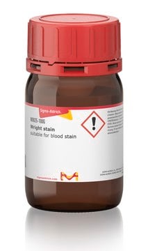
form
solution
Quality Level
shelf life
Expiry date on the label.
IVD
for in vitro diagnostic use
concentration
0.3%
pH
6.75-6.95 (25 °C)
application(s)
hematology
histology
storage temp.
room temp
Looking for similar products? Visit Product Comparison Guide
General description
Application
- Phagocytosis assay and puberty.
- Estrous cycle examinations.
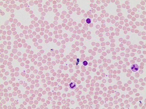
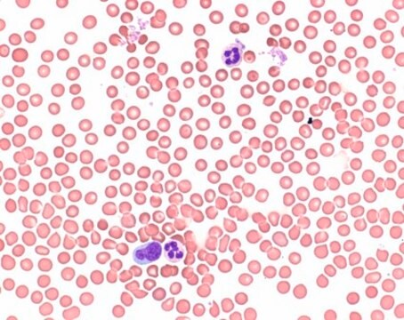
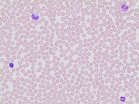
- Staining, to differentiate neutrophils.
Other Notes
Signal Word
Danger
Hazard Statements
Precautionary Statements
Hazard Classifications
Acute Tox. 3 Dermal - Acute Tox. 3 Inhalation - Acute Tox. 3 Oral - Eye Irrit. 2 - Flam. Liq. 3 - STOT SE 1
Target Organs
Eyes,Central nervous system
Storage Class Code
3 - Flammable liquids
WGK
WGK 2
Flash Point(F)
91.4 °F - closed cup
Flash Point(C)
33 °C - closed cup
Personal Protective Equipment
Certificates of Analysis (COA)
Search for Certificates of Analysis (COA) by entering the products Lot/Batch Number. Lot and Batch Numbers can be found on a product’s label following the words ‘Lot’ or ‘Batch’.
Already Own This Product?
Find documentation for the products that you have recently purchased in the Document Library.
Customers Also Viewed
Related Content
Learn about the clinical study of blood, blood-forming organs, and blood diseases including the treatment, prevention, and stains and dyes used in hematology testing.
Learn about the clinical study of blood, blood-forming organs, and blood diseases including the treatment, prevention, and stains and dyes used in hematology testing.
Learn about the clinical study of blood, blood-forming organs, and blood diseases including the treatment, prevention, and stains and dyes used in hematology testing.
Learn about the clinical study of blood, blood-forming organs, and blood diseases including the treatment, prevention, and stains and dyes used in hematology testing.
Our team of scientists has experience in all areas of research including Life Science, Material Science, Chemical Synthesis, Chromatography, Analytical and many others.
Contact Technical Service