B3111
ANTI-FLAG® M2 antibody, Mouse monoclonal
Clone M2, purified from hybridoma cell culture in bioreactor
Synonym(s):
Anti-ddddk, Anti-dykddddk, M2 clone ANTI-FLAG
Sign Into View Organizational & Contract Pricing
All Photos(7)
About This Item
UNSPSC Code:
12352203
NACRES:
NA.43
Recommended Products
biological source
mouse
antibody form
purified immunoglobulin (purified IgG1 subclass)
clone
M2, monoclonal
shelf life
4 yr
purified by
using Protein A
storage temp.
−20°C
General description
Monoclonal ANTI-FLAG M2 is a purified immunoglobulin, IgG1, monoclonal antibody, purified from culture supernatant of hybridoma cells, that binds to FLAG® fusion proteins. Unlike ANTI-FLAG M1 antibody, the M2 antibody will recognize the FLAG sequence at the N-terminus, Met-N-terminus, C-terminus, or at an internal site of FLAG fusion proteins. Monoclonal ANTI-FLAG M2 is useful for identification and capture of FLAG fusion proteins by common immunological procedures such as Western blots and immunoprecipitation. It is also useful for affinity purification of FLAG fusion proteins when bound to a solid support.
form: solution pH 7.4, containing 15 mM sodium azide
concentration: 3.0-5.0 mg/mL
form: solution pH 7.4, containing 15 mM sodium azide
concentration: 3.0-5.0 mg/mL
Application
IB, IF, IP, FACS, ELISA
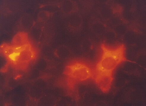
Antibody is recommended for use in several applications such as immunoblotting, immunoprecipitation, immunofluorescence, flow cytometry, and ELISA.
Learn more product details in our FLAG® application portal.
Antibody is recommended for use in several applications such as immunoblotting, immunoprecipitation, immunofluorescence, flow cytometry, and ELISA.
Learn more product details in our FLAG® application portal.
Packaging
polypropylene screw cap vial
Preparation Note
Dilute the antibody solution from 0.5-10 ug/mL in specified buffer
Legal Information
ANTI-FLAG is a registered trademark of Merck KGaA, Darmstadt, Germany
FLAG is a registered trademark of Merck KGaA, Darmstadt, Germany
Storage Class Code
12 - Non Combustible Liquids
WGK
nwg
Flash Point(F)
Not applicable
Flash Point(C)
Not applicable
Certificates of Analysis (COA)
Search for Certificates of Analysis (COA) by entering the products Lot/Batch Number. Lot and Batch Numbers can be found on a product’s label following the words ‘Lot’ or ‘Batch’.
Already Own This Product?
Find documentation for the products that you have recently purchased in the Document Library.
Runming Zeng et al.
Experimental biology and medicine (Maywood, N.J.), 246(6), 644-653 (2020-12-11)
Osteoarthritis (OA), the most prevalent form of arthritis disease, is characterized by destruction of articular cartilage, osteophyte development, and sclerosis of subchondral bone. Transcription factors Janus kinase 1/signal transducer and activator of transcription 3 (JAK1/STAT3) and Forkhead box M1 (FOXM1)
FBXO42 facilitates Notch signaling activation and global chromatin relaxation by promoting K63-linked polyubiquitination of RBPJ.
Jiang, et al.
Science Advances, 8, eabq4831-eabq4831 (2022)
Rui Santalla Méndez et al.
Cellular and molecular life sciences : CMLS, 80(10), 306-306 (2023-09-27)
Intracellular vesicle transport is essential for cellular homeostasis and is partially mediated by SNARE proteins. Endosomal trafficking to the plasma membrane ensures cytokine secretion in dendritic cells (DCs) and the initiation of immune responses. Despite its critical importance, the specific
Dongdong Shen et al.
PLoS pathogens, 20(7), e1012256-e1012256 (2024-07-18)
African swine fever (ASF) is a highly contagious, fatal disease of pigs caused by African swine fever virus (ASFV). The complexity of ASFV and our limited understanding of its interactions with the host have constrained the development of ASFV vaccines
Yi-Min Chu et al.
Frontiers in oncology, 12, 900166-900166 (2022-10-04)
DLC1 (deleted in liver cancer-1) is downregulated or deleted in colorectal cancer (CRC) tissues and functions as a potent tumor suppressor, but the underlying molecular mechanism remains elusive. We found that the conditioned medium (CM) collected from DLC1-overexpressed SW1116 cells
Our team of scientists has experience in all areas of research including Life Science, Material Science, Chemical Synthesis, Chromatography, Analytical and many others.
Contact Technical Service


