Skip to Content
Merck
All Photos(1)

Key Documents

C9598

Sigma-Aldrich

Anti-Caspase 3 antibody produced in rabbit

IgG fraction of antiserum, buffered aqueous solution

Synonym(s):

Anti-Apopain, Anti-CPP32, Anti-Yama

Sign Into View Organizational & Contract Pricing


About This Item

MDL number:
UNSPSC Code:
12352203
NACRES:
NA.46

biological source

rabbit

Quality Level

conjugate

unconjugated

antibody form

IgG fraction of antiserum

antibody product type

primary antibodies

clone

polyclonal

form

buffered aqueous solution

mol wt

antigen 32 kDa

species reactivity

human

technique(s)

microarray: suitable
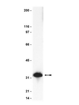
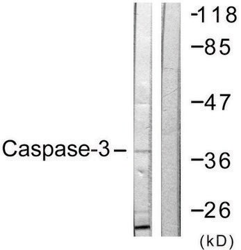
western blot: 1:3,000 using Jurkat human T-cell leukemia cell extract

UniProt accession no.

shipped in

dry ice

storage temp.

−20°C

target post-translational modification

unmodified

Gene Information

human ... CASP3(836)

General description

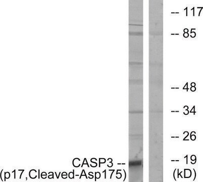
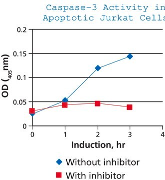
Caspase 3 is a cytosolic protein present as a 32 kDa proenzyme in cells. It is activated by proteolytic cleavage into the 20 kDa (p20) and 11 kDa (p11) active subunits when cells undergo apoptosis.

Immunogen

synthetic peptide corresponding to the N-terminal of human caspase 3 (amino acids 29-43 with C-terminally added lysine) conjugated to KLH. This sequence, unique to caspase 3, corresponds to N-terminal of the enzyme p20 subunit and is highly conserved in rat and mouse caspase 3.

Application

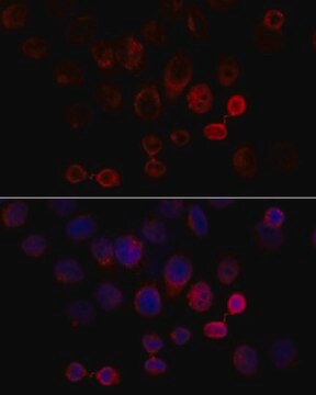
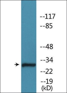
Anti-Caspase 3 antibody produced in rabbit is suitable for microarray and western blotting at a working dilution of 1:3000 using Jurkat human T-cell leukemia cell extract. It was used as a primary antibody for western blotting in a study to establish the biomolecular basis for the role of PCMT1 (isoaspartyl protein carboxyl-O-methyltransferase) as a part of a novel apoptosis regulatory mechanism. It was used to examine activation of caspase-3 via immunoblotting in a study to confirm the apoptosis of tumor cells mediated by AAV-ISN-T (recombinant adeno-associated virus vector with signal peptides of human insulin) administration. It was used as a primary antibody at 1:300 dilution for immunohistochemistry of deparaffinized sections of squamous cell carcinoma of the tongue containing surrounding normal tissue. It was used at a dilution of 1:1000 to detect caspase-3 and its cleaved form P20 in a study.

Biochem/physiol Actions

Caspase 3 cleaves many key proteins including poly(ADP-ribose) polymerase (PARP), sterol-regulatory element-binding proteins (SREBPs), DNA-dependent protein kinase (DNA-PK), α-fodrin, gelsolin, PKCd and DFF45/ICAD during apoptosis. High levels of this enzyme in lymphocytes suggest that it is an important mediator of apoptosis in the immune system. Deletion of CASP-3 gene in mice results in hyperplasia and cell abnormalities, indicating that caspase 3 is essential for morphogenetic cell death during normal brain development.
Caspase 3 is one of the key executioners of apoptosis downstream in the apoptotic pathway, as it is activated in cells by various death signals. In some neurodegenerative diseases, such as Huntington disease (HD) and Alzheimer′s disease (AD), specific neuronal caspase substrates have been identified. In Huntington disease (HD), caspase 3 specifically cleaves the HD gene product, Huntingtin.

Physical form

Solution in 0.01 M phosphate buffered saline, pH 7.4, containing 15 mM sodium azide.

Disclaimer

Unless otherwise stated in our catalog or other company documentation accompanying the product(s), our products are intended for research use only and are not to be used for any other purpose, which includes but is not limited to, unauthorized commercial uses, in vitro diagnostic uses, ex vivo or in vivo therapeutic uses or any type of consumption or application to humans or animals.

Not finding the right product?  

Try our Product Selector Tool.

Storage Class Code

10 - Combustible liquids

WGK

nwg

Flash Point(F)

Not applicable

Flash Point(C)

Not applicable


Certificates of Analysis (COA)

Search for Certificates of Analysis (COA) by entering the products Lot/Batch Number. Lot and Batch Numbers can be found on a product’s label following the words ‘Lot’ or ‘Batch’.

Already Own This Product?

Find documentation for the products that you have recently purchased in the Document Library.

Visit the Document Library

Alterations in Gene Expression Associated with Head and Neck Squamous Cell Carcinoma Development.
Serewko-Auret, Magdalena M
Cancer, Genomics, and Proteomics, 1, 137-148 (2004)
Nan Zhang et al.
Mediators of inflammation, 2014, 370530-370530 (2014-05-31)
The most effective way to contain cerebral ischemic injury is reperfusion; however, reperfusion itself may result in tissue injury, for which inflammatory damage is one of the main causative factors. NALP3 inflammasome is a multiprotein complex. It consists of NALP3
Q Song et al.
The EMBO journal, 15(13), 3238-3246 (1996-07-01)
Radiosensitive cell lines derived from X-ray cross complementing group 5 (XRCC5), SCID mice and a human glioma cell line lack components of the DNA-dependent protein kinase, DNA-PK, suggesting that DNA-PK plays an important role in DNA double-strand break repair. Another
Apoptosis: a review of programmed cell death.
Elmore S.
Toxicologic Pathology, 35(4), 495-516 (2007)
Hong Ma et al.
Hepatology (Baltimore, Md.), 42(6), 1355-1363 (2005-12-01)
The extracellular domain of the tumor necrosis factor-related apoptosis-inducing ligand (sTRAIL) may function as a soluble cytokine to selectively kill various cancer cells without toxicity to most normal cells. We constructed a series of recombinant adeno-associated virus (AAV) vectors expressing

Our team of scientists has experience in all areas of research including Life Science, Material Science, Chemical Synthesis, Chromatography, Analytical and many others.

Contact Technical Service