General description
Monoclonal Anti-Human IgG1 (mouse IgG2a isotype) is derived from the hybridoma produced by the fusion of mouse myeloma cells and splenocytes from an immunized mouse. Human IgG consists of four subclasses (1-4) that can be recognized by antigen differences in their heavy chains. They constitute approximately 65, 25, 6 and 4% of the total IgG, respectively. Each subclass has different biological and physiochemical properties.
The IgG subclass may be preferentially produced in response to different antigens. For instance, antipolysaccharide responses are mainly of the IgG2 subclass, while protein antigens give rise to IgG1 and IgG3 antibodies. Lipopolysaccahrides stimulate an IgG2 response in PBL′s and an IgG1 response in the spleen. Human IgG1 is the predominant subclass of in vivo and in vitro produced anti-tetanus toxoid antibodies. Only IgG1 and IgG3 are capable of adherence to mononuclear phagocytes. Serum IgG subclass deficiencies have been recorded for different patient groups. For example, IgG2 and IgG4 deficiency is associated with IgA deficiency as found in patients of ataxia telangiextasia. Low IgG2 levels were found in patients with SLE and juvenile diabetes melitus. A disproportionate elevation of IgG1 has also been found in the cerebral spinal fluid of patients with multiple sclerosis. Examination of the distribution pattern of IgG subclasses in different types of diseases may provide insight into the immunological processes involved and may assist in the diagnosis of various disorders.
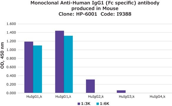
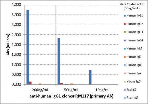
The antibody is specific for the Fc portion of human IgG1 and is non-reactive with other IgG subclasses. This clone, also described as HP-6091, was found to exhibit a valuable profile of reactivity and specificity for human IgG1 by the IUIS/WHO study.
Specificity
Human IgG consists of four subclasses (1-4) that can be recognized.
Application
Monoclonal Anti-Human IgG1 antibody produced in mouse has been used in:
- direct hemagglutination(HA)
- hemagglutination inhibition(HAI) assays
- enzyme linked immunosorbent assay (ELISA)
- immunofluorometric assay (IFMA)
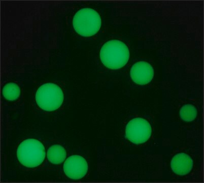
- immunofluorescence
- cell-based assays for the detection of IgG subclasses of acetylcholine receptor (AChR) antibodies
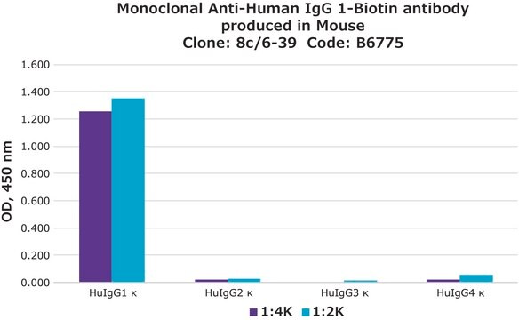
Mouse monoclonal clone clone 8c/6-39 anti-Human IgG1 antibody may be used for the identification of human IgG1 subclass by means of various immunoassays and direct hemagglutination (HA) and hemagglutination inhibition (HAI) assays, enzyme linked immunosorbent assay (ELISA), immunofluorometric assay (IFMA) and detection of cytoplasmic IgG. The antibody may be useful in the analysis of the distribution of IgG subclasses in various diseases.
Biochem/physiol Actions
Human IgG1 is the predominant subclass of in vivo and in vitro produced anti-tetanus toxoid antibodies. Only IgG1 and IgG3 are capable of adherence to mononuclear phagocytes via Fc receptors (FcR). A disproportionate elevation of IgG1 has also been found in the cerebral spinal fluid of patients with multiple sclerosis.
Physical form
The antibody is provided as ascites fluid with 0.1% sodium azide as a preservative.
Storage and Stability
For continuous use, store at 0-5 °C. For extended storage, the solution may be frozen in working aliquots. Repeated freezing and thawing is not recommended. Storage in "frost-free" freezers is not recommended. If slight turbidity occurs upon prolonged storage, clarify by centrifugation before use.
Disclaimer
Unless otherwise stated in our catalog or other company documentation accompanying the product(s), our products are intended for research use only and are not to be used for any other purpose, which includes but is not limited to, unauthorized commercial uses, in vitro diagnostic uses, ex vivo or in vivo therapeutic uses or any type of consumption or application to humans or animals.
