12013819001
Roche
Anti-HA-Peroxidase, High Affinity
from rat IgG1
Synonym(s):
antibody
Sign Into View Organizational & Contract Pricing
All Photos(1)
About This Item
UNSPSC Code:
12352203
Recommended Products
biological source
rat
Quality Level
conjugate
peroxidase conjugate
antibody form
purified immunoglobulin
antibody product type
primary antibodies
clone
clone 3F10, monoclonal
form
lyophilized (clear, colorless solution after reconstitution)
packaging
pkg of 25 U (25 μg)
manufacturer/tradename
Roche
isotype
IgG1
epitope sequence
YPYDVPDYA
storage temp.
2-8°C
Related Categories
General description
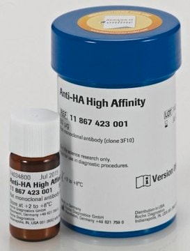
Anti-HA-Peroxidase, High Affinity is a monoclonal antibody to the HA-peptide (clone 3F10), conjugated to peroxidase.
Specificity
Anti-HA-Peroxidase, High Affinity (3F10) recognizes the 9-amino acid sequence YPYDVPDYA, derived from the human influenza hemagglutinin (HA) protein.
This epitope is also recognized in fusion proteins regardless of its position (N-terminal, C-terminal or internal).
This epitope is also recognized in fusion proteins regardless of its position (N-terminal, C-terminal or internal).
Immunogen
The epitope consists of amino acids 98-106 from the human influenza virus hemagglutinin protein.
Application
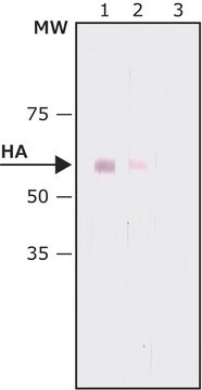
- Use Anti-HA-Peroxidase, High Affinity for the detection of HA-tagged recombinant proteins using: ELISA
- Western blot
Preparation Note
Stabilizers: proteinaceous stabilizers
Working concentration: The working concentration of conjugate depends on application and substrate.
The following concentrations should be taken as a guideline:
Working concentration: The working concentration of conjugate depends on application and substrate.
The following concentrations should be taken as a guideline:
- Dot blot: 50 mU/ml
- ELISA: 25 mU/ml
- Western blot: 50 mU/ml
Reconstitution
Add 1.0 ml double-distilled water for a final concentration of 25 U/mL.
Rehydrate for 10 min prior to use.
Rehydrate for 10 min prior to use.
Other Notes
For life science research only. Not for use in diagnostic procedures.
Not finding the right product?
Try our Product Selector Tool.
Signal Word
Warning
Hazard Statements
Precautionary Statements
Hazard Classifications
Skin Sens. 1
Storage Class Code
11 - Combustible Solids
WGK
WGK 1
Flash Point(F)
does not flash
Flash Point(C)
does not flash
Choose from one of the most recent versions:
Already Own This Product?
Find documentation for the products that you have recently purchased in the Document Library.
Customers Also Viewed
Regina Gratz et al.
The New phytologist, 225(1), 250-267 (2019-09-06)
The key basic helix-loop-helix (bHLH) transcription factor in iron (Fe) uptake, FER-LIKE IRON DEFICIENCY-INDUCED TRANSCRIPTION FACTOR (FIT), is controlled by multiple signaling pathways, important to adjust Fe acquisition to growth and environmental constraints. FIT protein exists in active and inactive
Ranran Wang et al.
Cancers, 12(6) (2020-06-25)
The epigenetic reader BRD4 binds acetylated histones and plays a central role in controlling cellular gene transcription and proliferation. Dysregulation of BRD4's activity has been implicated in the pathogenesis of a wide variety of cancers. While blocking BRD4 interaction with
Julia K Günther et al.
Biology, 9(5) (2020-05-21)
Rigosertib, via reactive oxygen species (ROS), stimulates cJun N-terminal kinases 1/2 (JNK1/2), which inactivate RAS/RAF signaling and thereby inhibit growth and survival of tumor cells. JNK1/2 are not only regulated by ROS-they in turn can also control ROS production. The
Satoshi Hashimoto et al.
Scientific reports, 10(1), 3422-3422 (2020-02-27)
Ribosome stalling triggers the ribosome-associated quality control (RQC) pathway, which targets collided ribosomes and leads to subunit dissociation, followed by proteasomal degradation of the nascent peptide. In yeast, RQC is triggered by Hel2-dependent ubiquitination of uS10, followed by subunit dissociation
Jihoon Ha et al.
International journal of molecular sciences, 21(9) (2020-05-02)
p62/sequestosome-1 is a scaffolding protein involved in diverse cellular processes such as autophagy, oxidative stress, cell survival and death. It has been identified to interact with atypical protein kinase Cs (aPKCs), linking these kinases to NF-κB activation by tumor necrosis
Our team of scientists has experience in all areas of research including Life Science, Material Science, Chemical Synthesis, Chromatography, Analytical and many others.
Contact Technical Service