Skip to Content
Merck
All Photos(1)

Key Documents

C8218

Sigma-Aldrich

Monoclonal Anti-Cdk4 antibody produced in mouse

clone DCS-31, ascites fluid

Sign Into View Organizational & Contract Pricing


About This Item

MDL number:
UNSPSC Code:
12352203
NACRES:
NA.41

biological source

mouse

conjugate

unconjugated

antibody form

ascites fluid

antibody product type

primary antibodies

clone

DCS-31, monoclonal

mol wt

antigen 33 kDa

contains

15 mM sodium azide

species reactivity

rat, mouse, human

technique(s)

immunocytochemistry: suitable
immunoprecipitation (IP): suitable
microarray: suitable
western blot: 1:1,000 using a cultured human tumor cell line extract

isotype

IgG2a

UniProt accession no.

shipped in

dry ice

storage temp.

−20°C

target post-translational modification

unmodified

Gene Information

human ... CDK4(1019)
mouse ... Cdk4(12567)
rat ... Cdk4(94201)

General description

Cdk4 exists, in part, as a multi-protein complex with a D-type cyclin, proliferating cell nuclear antigen (PCNA) and a protein inhibitor, p21Cip1. Cdk4 associates separately with p16, particularly in cells lacking a functional retinoblastoma protein.
Monoclonal Anti-Cdk4 (mouse IgG2a isotype) is derived from the DCS-31 hybridoma produced by the fusion of mouse myeloma cells and splenocytes from an immunized mouse. Cyclin-dependent kinase 4 is localized in human chromosome 12q14.1.

Specificity

The antibody reacts specifically with Cdk4 and does not recognize other Cdk types.

Immunogen

recombinant human Cdk4 protein

Application

Monoclonal Anti-Cdk4 antibody was used in:
  • immunoprecipitation of human melanoma cell line.
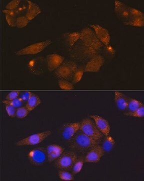
  • immunofluorescence of fibrosarcoma cells.
  • western blot analysis
  • immunofluorescence
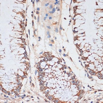
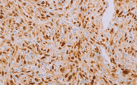
  • immunohistochemistry

Biochem/physiol Actions

In association with cyclins, cyclin-dependent kinases (CDKs) forms active kinase complexes which is responsible for regulating cell cycle progression in eukaryotic cells. Within the complexes, the CDKs serves a catalytic protein kinase activity. This catalytic activity is regulated by two mechanisms: protein phosphorylation and association of regulatory subunits.

Disclaimer

Unless otherwise stated in our catalog or other company documentation accompanying the product(s), our products are intended for research use only and are not to be used for any other purpose, which includes but is not limited to, unauthorized commercial uses, in vitro diagnostic uses, ex vivo or in vivo therapeutic uses or any type of consumption or application to humans or animals.

Not finding the right product?  

Try our Product Selector Tool.

Storage Class Code

10 - Combustible liquids

WGK

WGK 3

Flash Point(F)

Not applicable

Flash Point(C)

Not applicable


Certificates of Analysis (COA)

Search for Certificates of Analysis (COA) by entering the products Lot/Batch Number. Lot and Batch Numbers can be found on a product’s label following the words ‘Lot’ or ‘Batch’.

Already Own This Product?

Find documentation for the products that you have recently purchased in the Document Library.

Visit the Document Library

Adam Z Oskowitz et al.
The international journal of biochemistry & cell biology, 43(11), 1563-1572 (2011-07-29)
Recently we demonstrated that the miRNA regulate human mesenchymal stem cells (hMSCs) differentiation. To determine the role of the miRNA pathway in hMSCs proliferation, Drosha and Dicer knockdown hMSCs were generated using a lentiviral based tetracycline inducible shRNA. hMSCs with
HMGA2 is the partner of MDM2 in well-differentiated and dedifferentiated liposarcomas whereas CDK4 belongs to a distinct inconsistent amplicon
Italiano A, et al.
International Journal of Cancer. Journal International Du Cancer, 122(10), 2233-2241 (2008)
V Cecchinato et al.
Leukemia research, 32(5), 791-797 (2007-10-30)
T acute lymphoblastic leukemia cell lines treated with hexamethylene bisacetamide (HMBA) undergo a delay in cell cycle progression and increase susceptibility to apoptosis, although they never overcome the differentiation block. In accordance with changes in cell cycle and apoptosis, transitory
J Pines et al.
The Journal of cell biology, 115(1), 1-17 (1991-10-01)
We have used immunofluorescence staining to study the subcellular distribution of cyclin A and B1 during the somatic cell cycle. In both primary human fibroblasts and in epithelial tumor cells, we find that cyclin A is predominantly nuclear from S
Christoffer Bento et al.
BMC cell biology, 10, 89-89 (2009-12-19)
The DDIT3 gene encodes a transcription factor belonging to the CCAAT/enhancer binding protein (C/EBP) family. It is normally expressed at very low levels but is activated by cellular stress conditions and induces G1 arrest and, in some cell types, apoptosis.

Articles

Quantitative and qualitative western blotting to validate knockdown by esiRNA.

Our team of scientists has experience in all areas of research including Life Science, Material Science, Chemical Synthesis, Chromatography, Analytical and many others.

Contact Technical Service