Skip to Content
Merck
All Photos(6)

Key Documents

HPA001383

Sigma-Aldrich

Anti-ERBB2 antibody produced in rabbit

Ab1, Prestige Antibodies® Powered by Atlas Antibodies, affinity isolated antibody, buffered aqueous glycerol solution

Synonym(s):

Anti-CD340, Anti-HER-2, Anti-HER2, Anti-NEU, Anti-NGL, Anti-ERBB2

Sign Into View Organizational & Contract Pricing


About This Item

MDL number:
UNSPSC Code:
12352203
Human Protein Atlas Number:
NACRES:
NA.41

biological source

rabbit

Quality Level

conjugate

unconjugated

antibody form

affinity isolated antibody

antibody product type

primary antibodies

clone

polyclonal

product line

Prestige Antibodies® Powered by Atlas Antibodies

form

buffered aqueous glycerol solution

species reactivity

human

technique(s)

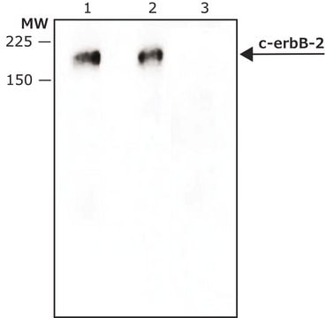
immunoblotting: 0.04-0.4 μg/mL
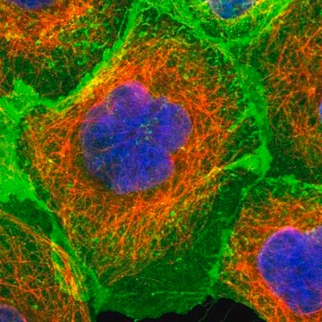
immunofluorescence: 0.25-2 μg/mL
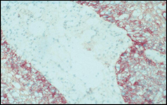
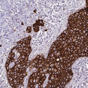
immunohistochemistry: 1:500-1:1000

immunogen sequence

LPSETDGYVAPLTCSPQPEYVNQPDVRPQPPSPREGPLPAARPAGATLERPKTLSPGKNGVVKDVFAFGGAVENPEYLTPQGGAAPQPHPPPAFSPAFDNLYYWDQDPPERGAPPSTFKGTPTA

UniProt accession no.

shipped in

wet ice

storage temp.

−20°C

target post-translational modification

unmodified

Gene Information

human ... ERBB2(2064)

Looking for similar products? Visit Product Comparison Guide

Immunogen

erb-b2 receptor tyrosine kinase 2

Application

Applications in which this antibody has been used successfully, and the associated peer-reviewed papers, are given below.
Western Blotting (1 paper)

Biochem/physiol Actions

Receptor tyrosine-protein kinase erbB-2 is a protein tyrosine kinase that is encoded by the gene ERBB2. It belongs to the epidermal growth factor (EGF) receptor family. The protein lacks a ligand binding domain and requires co-receptors for ligand binding. It functions as a transcriptional regulator by binding to cyclooxygenase enzyme COX-2 gene promoter. Overexpression of this gene has been observed in several types of cancer including breast and ovarian cancers. ErbB-2 enhances the EGF and NDF induced tyrosine phosphorylation of other ErbB family of receptor tyrosine kinases.

Features and Benefits

Prestige Antibodies® are highly characterized and extensively validated antibodies with the added benefit of all available characterization data for each target being accessible via the Human Protein Atlas portal linked just below the product name at the top of this page. The uniqueness and low cross-reactivity of the Prestige Antibodies® to other proteins are due to a thorough selection of antigen regions, affinity purification, and stringent selection. Prestige antigen controls are available for every corresponding Prestige Antibody and can be found in the linkage section.

Every Prestige Antibody is tested in the following ways:
  • IHC tissue array of 44 normal human tissues and 20 of the most common cancer type tissues.
  • Protein array of 364 human recombinant protein fragments.

Linkage

Corresponding Antigen APREST79717

Physical form

Solution in phosphate-buffered saline, pH 7.2, containing 40% glycerol and 0.02% sodium azide

Legal Information

Prestige Antibodies is a registered trademark of Merck KGaA, Darmstadt, Germany

Disclaimer

Unless otherwise stated in our catalog or other company documentation accompanying the product(s), our products are intended for research use only and are not to be used for any other purpose, which includes but is not limited to, unauthorized commercial uses, in vitro diagnostic uses, ex vivo or in vivo therapeutic uses or any type of consumption or application to humans or animals.

Not finding the right product?  

Try our Product Selector Tool.

Storage Class Code

10 - Combustible liquids

WGK

WGK 1

Flash Point(F)

Not applicable

Flash Point(C)

Not applicable

Personal Protective Equipment

dust mask type N95 (US), Eyeshields, Gloves

Certificates of Analysis (COA)

Search for Certificates of Analysis (COA) by entering the products Lot/Batch Number. Lot and Batch Numbers can be found on a product’s label following the words ‘Lot’ or ‘Batch’.

Already Own This Product?

Find documentation for the products that you have recently purchased in the Document Library.

Visit the Document Library

Hee Jin Lee et al.
American journal of clinical pathology, 142(6), 755-766 (2014-11-13)
Heterogeneity of HER2 gene amplification is found in a subset of breast cancers. We investigated the impact of HER2 heterogeneity on trastuzumab responses and clinical outcomes in 112 patients with HER2-positive metastatic breast cancer. Regional and genetic heterogeneity of HER2
Xin-Zu Chen et al.
Hepato-gastroenterology, 61(134), 1817-1821 (2014-12-02)
The aim of this study was to investigate the association of human epidermal growth factor receptor 2 (HER2) expression with clinicopathological characteristics of resectable gastric cancer patients. A total of 394 stage I-III surgical gastric cancer patients who were detected
R Vadlamudi et al.
Oncogene, 18(2), 305-314 (1999-02-02)
Emerging lines of evidence suggest that in addition to growth factors, the process of colorectal tumorigenesis may also be driven by the upregulation of the inducible form of cyclooxygenase-2 (COX-2), an enzyme responsible for the conversion of arachidonic acid to
Lotte K Kristensen et al.
Theranostics, 9(15), 4409-4420 (2019-07-10)
Antibody-based PET tracers are exceptionally well-suited for determination of the in vivo biodistribution and quantification of therapeutic antibodies. The continued expansion in antibody-based therapeutics has accordingly driven the development towards more robust conjugation strategies in order to reliably predict the
Gail M Seigel et al.
Oncoscience, 6(7-8), 357-366 (2019-10-15)
Retinoblastoma (RB) is an ocular tumor of early childhood. Current treatments attempt to preserve visual function, but may spare chemoresistant tumor cells. One potential therapeutic target for RB is HER2, (ERBB2), expressed in RB in truncated form. In this study

Our team of scientists has experience in all areas of research including Life Science, Material Science, Chemical Synthesis, Chromatography, Analytical and many others.

Contact Technical Service