Skip to Content
Merck
All Photos(12)

Key Documents

V9131

Sigma-Aldrich

Anti-Vinculin Antibody

enhanced validation

mouse monoclonal, hVIN-1

Synonym(s):

Anti Vinculin, Vinculin Antibody, Vinculin Antibody - Monoclonal Anti-Vinculin antibody produced in mouse

Sign Into View Organizational & Contract Pricing


About This Item

MDL number:
UNSPSC Code:
12352203
NACRES:
NA.41

product name

Monoclonal Anti-Vinculin antibody produced in mouse, clone hVIN-1, ascites fluid

biological source

mouse

Quality Level

conjugate

unconjugated

antibody form

ascites fluid

antibody product type

primary antibodies

clone

hVIN-1, monoclonal

mol wt

antigen 116 kDa

contains

15 mM sodium azide

species reactivity

frog, chicken, mouse, canine, human, bovine, rat, turkey

enhanced validation

independent
Learn more about Antibody Enhanced Validation

technique(s)

immunohistochemistry (frozen sections): suitable
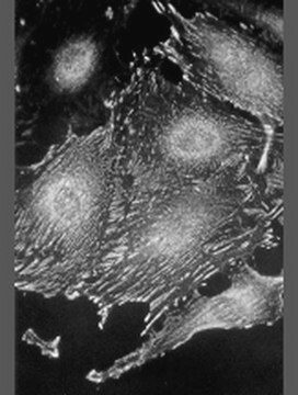
indirect immunofluorescence: 1:400 using cultured human fibroblasts
western blot: 1:200 using extract of human fibroblasts

isotype

IgG1

UniProt accession no.

application(s)

research pathology

shipped in

dry ice

storage temp.

−20°C

target post-translational modification

unmodified

Gene Information

human ... VCL(7414)
mouse ... Vcl(22330)
rat ... Vcl(305679)

General description

Monoclonal Anti-Vinculin (mouse IgG1 isotype) is derived from the hVIN-1 hybridoma produced by the fusion of mouse myeloma cells and splenocytes from immunized BALB/c mice. Vinculin is localized in the fascia adherens of the intercalated disk (cardiac muscle), myotendinous junctions (skeletal muscle), neuromuscular junctions and the membrane-associated dense bodies of smooth muscle. In many cell types undergoing viral transformation, vinculin becomes redistributed to rosettes or podosomes.

Specificity

Monoclonal Anti-Vinculin specifically stains vinculin at cell-cell and cell-substrate contacts in tissue and cultured cells using indirect immunofluorescent labeling. The antibody reacts with the 116 kDa vinculin band in immunoblotting. The product reacts with vinculin of many species. Good reactivity is obtained with human, bovine, chicken, dog, rat, mouse, turkey, and Xenopus. The antibody shows cross reactivity with smooth muscle metavinculin.
Specifically labels vinculin at cell-cell and cell-substrate contacts. Reacts strongly with human vinculin. Shows cross-reactivity with smooth muscle metavinculin.

Immunogen

purified human vinculin from uterus.

Application

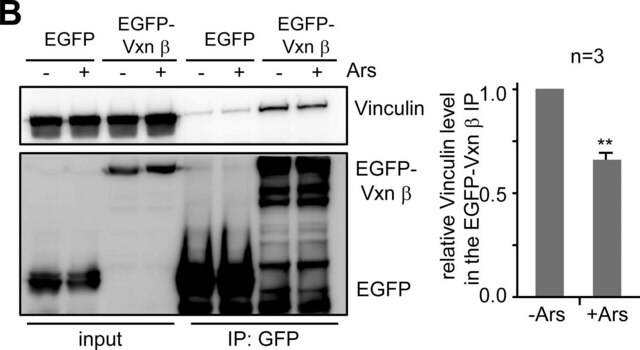
Monoclonal Anti-Vinculin antibody produced in mouse has been used in:
  • western blotting
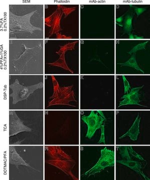
  • immunofluorescence staining
  • immunohistochemistry

Biochem/physiol Actions

Vinculin is a cytoskeletal protein associated with the cytoplasmic faces of both cell-cell and cell-extracellular matrix adherens-type junctions. It functions as one of several interacting proteins involved in anchoring F-actin to the membrane. A sequence of molecular interactions might be involved in the transmembrane assembly of adhesion plaques. In the assembly of adhesion plaques, the β-subunit of integrin binds to talin. Talin binds to vinculin that interacts with α-actinin and possibly with itself. Since α-actinin binds to and cross-links actin filaments, vinculin represents a key element in the transmembrane linkage of the extracellular matrix to the cytoplasmic microfilament system.

Disclaimer

Unless otherwise stated in our catalog or other company documentation accompanying the product(s), our products are intended for research use only and are not to be used for any other purpose, which includes but is not limited to, unauthorized commercial uses, in vitro diagnostic uses, ex vivo or in vivo therapeutic uses or any type of consumption or application to humans or animals.

Not finding the right product?  

Try our Product Selector Tool.

Storage Class Code

10 - Combustible liquids

WGK

WGK 3

Flash Point(F)

Not applicable

Flash Point(C)

Not applicable


Certificates of Analysis (COA)

Search for Certificates of Analysis (COA) by entering the products Lot/Batch Number. Lot and Batch Numbers can be found on a product’s label following the words ‘Lot’ or ‘Batch’.

Already Own This Product?

Find documentation for the products that you have recently purchased in the Document Library.

Visit the Document Library

Reinforcement versus fluidization in cytoskeletal mechanoresponsiveness
Krishnan R, et al.
PLoS ONE, 4(5), e5486-e5486 (2009)
Dystrophin-glycoprotein complex and vinculin-talin-integrin system in human adult cardiac muscle
Anastasi G, et al.
International Journal of Molecular Medicine, 23(2), 149-159 (2009)
Vinculin and talin: focus on the myocardium
Zemljic-Harpf A, et al.
Journal of Investigative Medicine, 57(8), 849-855 (2009)
Somatic inactivation of the PHD2 prolyl hydroxylase causes polycythemia and congestive heart failure
Minamishima YA, et al.
Blood, 111(6), 3236-3244 (2008)
Prognostic significance of epidermal growth factor receptor phosphorylation and mutation in head and neck squamous cell carcinoma
Hama T, et al.
Oncologist, 14(9), 900-908 (2009)

Articles

In the midst of beeping lab timers, presentations and grant deadlines, it is easy to take for granted the quality of lab reagents.

Learn differences in monoclonal vs polyclonal antibodies including how antibodies are generated, clone numbers, and antibody formats.

Immunofluorescence uses antibody-conjugated fluorescent molecules for protein localization, modification confirmation, and protein complex visualization.

Explore the basics of working with antibodies including technical information on structure, classes, and normal immunoglobulin ranges.

See All

Protocols

Tips and troubleshooting for FFPE and frozen tissue immunohistochemistry (IHC) protocols using both brightfield analysis of chromogenic detection and fluorescent microscopy.

Our team of scientists has experience in all areas of research including Life Science, Material Science, Chemical Synthesis, Chromatography, Analytical and many others.

Contact Technical Service