Skip to Content
Merck
All Photos(6)

Documents

MAB374

Sigma-Aldrich

Anti-Glyceraldehyde-3-Phosphate Dehydrogenase Antibody, clone 6C5

clone 6C5, Chemicon®, from mouse

Synonym(s):

glyceraldehyde 3-phosphate dehydrogenase, aging-associated gene 9 protein, glyceraldehyde-3-phosphate dehydrogenase, GAPDH, G3PDH, GAPD, OK/SW-cl.12, MGC88685, G3PD, EC 1.2.1.12

Sign Into View Organizational & Contract Pricing


About This Item

UNSPSC Code:
12352203
eCl@ss:
32160702
NACRES:
NA.41

biological source

mouse

Quality Level

antibody form

purified immunoglobulin

antibody product type

primary antibodies

clone

6C5, monoclonal

species reactivity

human, feline, pig, mouse, rabbit, fish, canine, rat

should not react with

E. coli

manufacturer/tradename

Chemicon®

technique(s)

ELISA: suitable
immunocytochemistry: suitable
immunofluorescence: suitable
immunohistochemistry: suitable
immunoprecipitation (IP): suitable
western blot: suitable

isotype

IgG1

NCBI accession no.

UniProt accession no.

shipped in

wet ice

target post-translational modification

unmodified

Gene Information

human ... GAPDH(2597)
mouse ... Gapdh(14433)

General description

Glyceraldehyde 3-phosphate dehydrogenase (GAPDH) is an ubiquitous glycolytic enzyme present in reasonably high levels in almost all tissues. As a ′house-keeping′ enzyme, it catalyzes the synthesis of 1,3-biphosphoglycerate, a "high energy" intermediate used for the synthesis of ATP. Besides its cytoplasmic action in metabolism it is also involved in the initial stages of apoptosis or oxidative stress response where GAPDH is translocated to the nucleus. Such actions may reflect the role of GAPDH in DNA repair or as one nuclear carrier for apoptotic molecules. GAPDH has also been found to bind specifically to proteins implicated in the pathogenesis of a variety of neurodegenerative disorders including the beta-amyloid precursor protein and the huntingtin protein where decreased function of GAPDH is associated with Alzheimer′s and Huntington′s disease fibroblasts. GAPDH has also been identified as a potential target for nitric oxide (NO)-mediated cellular toxicity. The complete and functional enzyme is a tetramer with each of four identical subunits occupying the vertex of a tetrahedron. Binding domains also include one principally interacting with NAD+ and another interacting with glyceraldehyde 3-phosphate (GAP).

Specificity

Glyceraldehyde-3-phosphate dehydrogenase (GAPDH) from skeletal muscle. Antibody also recognizes cardiac GAPDH. GAPDH enzyme is detected in many non-muscle cells lines including HeLa, HCT-116 cells, U937 and THP-1 cells among others.
Human, porcine, canine, rat, mouse, rabbit, cat, and fish skeletal muscle. It has been reported that this antibody does not react with GAPDH from E. coli.

Immunogen

Glyceraldehyde-3-phosphate dehydrogenase from rabbit muscle.

Application

Anti-Glyceraldehyde-3-Phosphate Dehydrogenase Antibody, clone 6C5 is a well published and extensively characterized monoclonal antibody. This purified mAb detects Glyceraldehyde-3-Phosphate Dehydrogenase (GAPDH) & has been published & validated for use in ELISA, IP, IC, IF, IH & WB.
ELISA:
1:1,000,000 dilution of a previous lot ws used for Immunoaffinity purification of GAPDH.

Immunocytochemistry:
1:100 to 1:300 dilution of a previous lot was used in immunocytochemistry in PBS-BSA 10 mg/mL. 4% PFA, with 0.02% PBS-Triton X-100; 3 min RT (Griffoni, 2001).

Western blot:
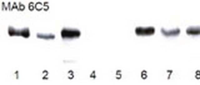
1:100 to 1:300. Recognizes a 36kDa band of the reduced monomer. Non-reduced GAPDH runs as a 146kDa tetramer.

Immunoaffinity purification of GAPDH.

Optimal working dilutions must be determined by end user.
Research Category
Metabolism
Research Sub Category
Enzymes & Biochemistry

Quality

Evaluated by Western Blot on A431 lysates.

Western Blot Analysis:
1:500 dilution of this antibody detected GLYCERALDEHYDE-3-PDH on 10 μg of A431 lysates.

Target description

38 kDa

Physical form

Format: Purified
Protein A purified
Purified Mouse monoclonal IgG1 in PBS, pH 7.4 with 0.1% sodium azide.

Storage and Stability

Maintain at 2-8ºC in undiluted aliquots for up to 6 months after date of receipt.

Analysis Note

Control
It is expressed in all cells.

Legal Information

CHEMICON is a registered trademark of Merck KGaA, Darmstadt, Germany

Disclaimer

Unless otherwise stated in our catalog or other company documentation accompanying the product(s), our products are intended for research use only and are not to be used for any other purpose, which includes but is not limited to, unauthorized commercial uses, in vitro diagnostic uses, ex vivo or in vivo therapeutic uses or any type of consumption or application to humans or animals.

Not finding the right product?  

Try our Product Selector Tool.

Storage Class Code

12 - Non Combustible Liquids

WGK

WGK 2

Flash Point(F)

Not applicable

Flash Point(C)

Not applicable


Certificates of Analysis (COA)

Search for Certificates of Analysis (COA) by entering the products Lot/Batch Number. Lot and Batch Numbers can be found on a product’s label following the words ‘Lot’ or ‘Batch’.

Already Own This Product?

Find documentation for the products that you have recently purchased in the Document Library.

Visit the Document Library

Studying protein degradation pathways in vivo using a cranial window-based approach.
Unni, VK; Ebrahimi-Fakhari, D; Vanderburg, CR; McLean, PJ; Hyman, BT
Methods null
Manoj B Menon et al.
PloS one, 6(8), e23054-e23054 (2011-08-20)
SB202190, a widely used inhibitor of p38 MAPKα and β, was recently described to induce autophagic vacuoles and cell death in colon and ovarian cancer cells lines and, therefore, this effect was supposed to be specific for transformed cells and
Kristina Leuner et al.
PloS one, 6(2), e14716-e14716 (2011-03-03)
Psoriasis is a characteristic inflammatory and scaly skin condition with typical histopathological features including increased proliferation and hampered differentiation of keratinocytes. The activation of innate and adaptive inflammatory cellular immune responses is considered to be the main trigger factor of
Anti-VEGF antibody treatment accelerates polycystic kidney disease.
Raina, S; Honer, M; Kramer, SD; Liu, Y; Wang, X; Segerer, S; Wuthrich, RP; Serra, AL
American Journal of Physiology: Renal Physiology null
An integrated systems analysis implicates EGR1 downregulation in simian immunodeficiency virus encephalitis-induced neural dysfunction.
Gersten, M; Alirezaei, M; Marcondes, MC; Flynn, C; Ravasi, T; Ideker, T; Fox, HS
The Journal of Neuroscience null

Articles

Immunofluorescence uses antibody-conjugated fluorescent molecules for protein localization, modification confirmation, and protein complex visualization.

Protocols

Tips and troubleshooting for FFPE and frozen tissue immunohistochemistry (IHC) protocols using both brightfield analysis of chromogenic detection and fluorescent microscopy.

Our team of scientists has experience in all areas of research including Life Science, Material Science, Chemical Synthesis, Chromatography, Analytical and many others.

Contact Technical Service