
04511
Live/Dead Cell Double Staining Kit
suitable for fluorescence
Synonym(s):
Staining kit for live/dead cells
Sign Into View Organizational & Contract Pricing
All Photos(1)
About This Item
UNSPSC Code:
12161503
NACRES:
NA.32
Recommended Products


Related Categories
Application
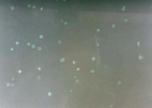
The Live/Dead Cell Double Staining Kit is utilized for simultaneous fluorescence staining of viable and dead cells. This kit contains calcein-AM and propidium iodide (PI) solutions, which stain viable and dead cells, respectively. Calcein-AM, acetoxymethyl ester of calcein, is highly lipophilic and cell membrane permeable. Though calcein-AM itself is not a fluorescent molecule, the calcein generated from Calcein-AM by esterase in a viable cell emits a strong green fluorescence (λex 490 nm, λem 515 nm). Therefore, calcein-AM only stains viable cells. Alternatively, the nuclei staining dye PI cannot pass through a viable cell membrane. It reaches the nucleus by passing through disordered areas of dead cell membrane, and intercalates with the DNA double helix of the cell to emit red fluorescence (λex 535 nm, λem 617 nm). Since both calcein and PI-DNA can be excited with 490 nm light, simultaneous monitoring of viable and dead cells is possible with a fluorescence microscope. Using λex 545 nm, only dead cells can be observed.
Kit Components Only
Product No.
Description
- Solution A (Calcein AM solution) 4 × 50
- Solution B (propidium iodide solution) 300 μL
related product
Product No.
Description
Pricing
Storage Class Code
10 - Combustible liquids
WGK
WGK 2
Flash Point(F)
185.0 °F - closed cup
Flash Point(C)
85 °C - closed cup
Choose from one of the most recent versions:
Already Own This Product?
Find documentation for the products that you have recently purchased in the Document Library.
Customers Also Viewed


T Kimura et al.
Neuroscience letters, 208(1), 53-56 (1996-04-12)
The recent immunological demonstration of advanced glycation end products (AGE) of the Maillard reaction in several human tissues suggests a possible involvement of AGE in the aging process. We previously prepared a monoclonal anti-AGE antibody (6D12) which recognized N epsilon-(carboxymethyl)lysine.
Zhen Cao et al.
Cellular and molecular neurobiology, 35(8), 1073-1079 (2015-07-03)
Scorpion venom has been used in the Orient to treat central nervous system diseases for many years, and the protein/peptide toxins in Buthus martensii Karsch (BmK) venom are believed to be the effective components. Scorpion venom heat-resistant peptide (SVHRP) is
Bleaching of plasmon-resonance absorption of gold nanorods decreases efficiency of cell destruction.
Florian Rudnitzki et al.
Journal of biomedical optics, 17(5), 058003-058003 (2012-05-23)
When irradiated with nanosecond laser pulses, gold nanoparticles allow for manipulation or destruction of cells and proteins with high spatial and temporal precision. Gold nanorods are especially attractive, because they have an up-to-20-fold stronger absorption than a sphere of equal
S Yoshida et al.
Clinical nephrology, 49(5), 273-280 (1998-06-09)
Cardiovascular disease is one of the most common complications of dialysis and renal transplant patients, and high levels of AGE are present in end-stage renal failure. To address the potential involvement of AGE and growth factors in the pathophysiology of
Amin Hassanzadeh-Barforoushi et al.
Lab on a chip, 18(15), 2156-2166 (2018-06-21)
We present here a new method to easily and reliably generate an array of hundreds of dispersed nanoliter-volume semi-droplets for single-cells culture and analysis. The liquid segmentation step occurs directly in indexed traps by a tweezer-like mechanism and is stabilized
Articles
Cell based assays for cell proliferation (BrdU, MTT, WST1), cell viability and cytotoxicity experiments for applications in cancer, neuroscience and stem cell research.
Our team of scientists has experience in all areas of research including Life Science, Material Science, Chemical Synthesis, Chromatography, Analytical and many others.
Contact Technical Service