Skip to Content
Merck
All Photos(1)

Key Documents

102M-1

Sigma-Aldrich

CD2 (MRQ-11) Mouse Monoclonal Antibody

Sign Into View Organizational & Contract Pricing


About This Item

UNSPSC Code:
12352203
NACRES:
NA.41

biological source

mouse

Quality Level

100
500

conjugate

unconjugated

antibody form

culture supernatant

antibody product type

primary antibodies

clone

MRQ-11, monoclonal

description

For In Vitro Diagnostic Use in Select Regions (See Chart)

form

buffered aqueous solution

species reactivity

human

packaging

vial of 0.1 mL concentrate (102M-14)
vial of 0.5 mL concentrate (102M-15)
bottle of 1.0 mL predilute (102M-17)
vial of 1.0 mL concentrate (102M-16)
bottle of 7.0 mL predilute (102M-18)

manufacturer/tradename

Cell Marque

IVD

for in vitro diagnostic use

technique(s)

immunohistochemistry (formalin-fixed, paraffin-embedded sections): 1:10-1:50 (concentrated)

isotype

IgG1κ

control

tonsil

shipped in

wet ice

storage temp.

2-8°C

visualization

cytoplasmic, membranous

Gene Information

human ... CD2(914)

General description

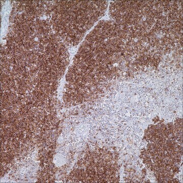
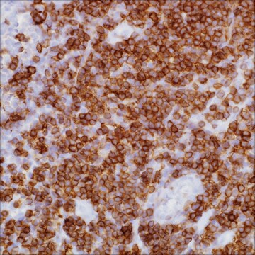
CD2 is a surface antigen present in all peripheral T-cell lymphocytes. CD2 is associated with signaling and mediating adhesion to other T-cells through the lymphocyte function-associated antigen and CD48/BCM1 complex. Anti-CD2 is a useful immunohistochemical reagent for the identification of normal T-lymphocytes, and for assessing lymphoid malignancies.

Quality


IVD

IVD

IVD

RUO

Linkage

CD2 Positive Control Slides, Product No. 102S, are available for immunohistochemistry (formalin-fixed, paraffin-embedded sections).

Physical form

Solution in Tris Buffer, pH 7.3-7.7, with 1% BSA and <0.1% Sodium Azide

Preparation Note

Download the IFU specific to your product lot and formatNote: This requires a keycode which can be found on your packaging or product label.

Other Notes

For Technical Service please contact: 800-665-7284 or email: service@cellmarque.com

Legal Information

Cell Marque is a trademark of Merck KGaA, Darmstadt, Germany

Not finding the right product?  

Try our Product Selector Tool.


Certificates of Analysis (COA)

Search for Certificates of Analysis (COA) by entering the products Lot/Batch Number. Lot and Batch Numbers can be found on a product’s label following the words ‘Lot’ or ‘Batch’.

Already Own This Product?

Find documentation for the products that you have recently purchased in the Document Library.

Visit the Document Library

L González et al.
Journal of comparative pathology, 125(1), 41-47 (2001-07-05)
This report describes a method (fixation, paraffin wax-embedding and immunolabelling) for the demonstration of several immune system cell epitopes (CD1, CD2, CD4, CD8, CD14, CD21, CD45R, WC-1, 28 kDa surface antigen, immunoglobulins and MHC II antigens) in ovine lymph nodes
H J Bovenschen et al.
The British journal of dermatology, 153(1), 72-78 (2005-07-21)
T-cell infiltration in plaque psoriasis has recently been an important subject of investigation. Interestingly, comparative analyses of the disease-specific composition of the lesional T-cell infiltrate in plaque psoriasis and other inflammatory dermatoses have only sparsely been performed. To compare plaque
Carlos Barrionuevo et al.
Applied immunohistochemistry & molecular morphology : AIMM, 15(1), 38-44 (2007-06-01)
It is well known that extranodal NK/T-cell lymphoma (NK/TCL) nasal type clusters in Asian countries. A large series of 78 cases of nasal NK/TCL from Peru is analyzed in the present study. Two histologic groups 1 (monomorphic) and 2 (polymorphic)
K A Foon et al.
Blood, 68(1), 1-31 (1986-07-01)
Important insights into leukocyte differentiation and the cellular origins of leukemia and lymphoma have been gained through the use of monoclonal antibodies that define cell surface antigens and molecular probes that identify immunoglobulin and T cell receptor genes. Results of
Nadine S I Aguilera et al.
Archives of pathology & laboratory medicine, 130(12), 1772-1779 (2006-12-08)
Reed-Sternberg cells in classic Hodgkin lymphoma are enigmatic and difficult to study because they are so sparse. Tissue microdissection allows for the isolation of single Reed-Sternberg cells. Isolated Reed-Sternberg cells show clonal immunoglobulin gene rearrangement indicating a B-cell origin. Rarely

Our team of scientists has experience in all areas of research including Life Science, Material Science, Chemical Synthesis, Chromatography, Analytical and many others.

Contact Technical Service