Skip to Content
Merck
All Photos(2)

Key Documents

D3320

Sigma-Aldrich

Anti-DVL1 (C-terminal) antibody produced in rabbit

enhanced validation

~1.5 mg/mL, affinity isolated antibody, buffered aqueous solution

Synonym(s):

Anti-Dishevelled-1, Anti-Dsh homolog 1, Anti-Segment polarity protein dishevelled homolog

Sign Into View Organizational & Contract Pricing


About This Item

UNSPSC Code:
12352203
NACRES:
NA.41

biological source

rabbit

Quality Level

conjugate

unconjugated

antibody form

affinity isolated antibody

antibody product type

primary antibodies

clone

polyclonal

form

buffered aqueous solution

mol wt

antigen ~85 kDa

species reactivity

human

enhanced validation

recombinant expression
Learn more about Antibody Enhanced Validation

concentration

~1.5 mg/mL

technique(s)

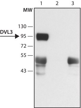
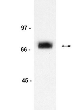
western blot: 2.0-4.0 μg/mL using HEK-293T cell lysate expressing human DVL1

UniProt accession no.

shipped in

dry ice

storage temp.

−20°C

target post-translational modification

unmodified

Gene Information

human ... DVL1(1855)

General description

The three DVL isoforms display high sequence homology and have conserved DIX, PDZ and DEP domains required for GSK3β inactivation. DVL1 is highly expressed in adult and fetal tissues such as skeletal muscle and pancreas but is also found in the brain and neural tube.

Application

Anti-DVL1 (C-terminal) antibody produced in rabbit has been used in immunoblotting and immunofluorescence.

Biochem/physiol Actions

Dishevelled (Dsh, DVL) operates by up-regulating β-catenin levels and stimulating T-cell factor (TCF)/lymphoid enhancer-binding factor 1 (LEF-1)-dependent transcription. In mammals, three genes encoding isoforms of dishevelled are present, DVL1, DVL2, and DVL3, that differentially mediate the wingless (wnt) canonical signaling pathway.
Dishevelled (Dsh, DVL) proteins are part of a multigene family that mediate wnt signaling pathways that are essential for the regulation of cellular proliferation, differentiation, motility, and morphogenesis. DVL1 regulates the activity of JNK (c-Jun N-terminal kinase) and GSK3β in the wnt signaling pathway. Upon activation of the wnt signaling pathway, DVL1 inactivates GSK3β through complex formation with APC (Antigen-presenting cell), β-catenin and axin proteins, releasing β-catenin from degradation. In neurons, DVL1 is localized to axonal microtubules and stabilizes microtubules by inhibiting the GSK3β mediated phosphorylation of MAP-1B.

Physical form

Solution in 0.01 M phos­phate buffered saline, pH 7.4, containing 15 mM sodium azide.

Disclaimer

Unless otherwise stated in our catalog or other company documentation accompanying the product(s), our products are intended for research use only and are not to be used for any other purpose, which includes but is not limited to, unauthorized commercial uses, in vitro diagnostic uses, ex vivo or in vivo therapeutic uses or any type of consumption or application to humans or animals.

Not finding the right product?  

Try our Product Selector Tool.

Storage Class Code

10 - Combustible liquids

Flash Point(F)

Not applicable

Flash Point(C)

Not applicable

Personal Protective Equipment

dust mask type N95 (US), Eyeshields, Gloves

Choose from one of the most recent versions:

Certificates of Analysis (COA)

Lot/Batch Number

Don't see the Right Version?

If you require a particular version, you can look up a specific certificate by the Lot or Batch number.

Already Own This Product?

Find documentation for the products that you have recently purchased in the Document Library.

Visit the Document Library

Exosomes mediate stromal mobilization of autocrine Wnt-PCP signaling in breast cancer cell migration
Luga V, et al.
Cell, 151(7), 1542-1556 (2012)
Rac1 acts in conjunction with Nedd4 and dishevelled-1 to promote maturation of cell-cell contacts
Nethe M, et al.
Journal of Cell Science, 125(14), 3430-3442 (2012)
L Li et al.
The Journal of biological chemistry, 274(1), 129-134 (1998-12-29)
Dishevelled (Dsh/Dvl) proteins are known to mediate Wnt signaling by up-regulating beta-catenin levels and stimulating T cell factor (TCF)/LEF-1-dependent transcription. We have identified a new Dvl-mediated signaling pathway in that mouse Dvl proteins, when expressed in COS-7 cells, stimulate c-Jun-dependent
Lorenza Ciani et al.
The Journal of cell biology, 164(2), 243-253 (2004-01-22)
Dishevelled (DVL) is associated with axonal microtubules and regulates microtubule stability through the inhibition of the serine/threonine kinase, glycogen synthase kinase 3beta (GSK-3beta). In the canonical WNT pathway, the negative regulator Axin forms a complex with beta-catenin and GSK-3beta, resulting
Petra Paclíková et al.
Molecular and cellular biology, 37(18) (2017-07-05)
Dishevelled (DVL) proteins are key mediators of the Wnt/β-catenin signaling pathway. All DVL proteins contain three conserved domains: DIX, PDZ, and DEP. There is a consensus in the field that the DIX domain is critical for Wnt/β-catenin signaling, but contradictory

Our team of scientists has experience in all areas of research including Life Science, Material Science, Chemical Synthesis, Chromatography, Analytical and many others.

Contact Technical Service