Skip to Content
Merck
All Photos(3)

Key Documents

ABE542

Sigma-Aldrich

Anti-YTHDF2 Antibody

from rabbit, purified by affinity chromatography

Synonym(s):

YTH domain-containing family protein 2, CLL-associated antigen KW-14, High-glucose-regulated protein 8, Renal carcinoma antigen NY-REN-2

Sign Into View Organizational & Contract Pricing


About This Item

UNSPSC Code:
12352203
eCl@ss:
32160702
NACRES:
NA.41
clone:
polyclonal
application:
ICC
WB
species reactivity:
human, rat
technique(s):
immunocytochemistry: suitable
western blot: suitable
citations:
9

biological source

rabbit

Quality Level

antibody form

affinity isolated antibody

antibody product type

primary antibodies

clone

polyclonal

purified by

affinity chromatography

species reactivity

human, rat

technique(s)

immunocytochemistry: suitable
western blot: suitable

NCBI accession no.

UniProt accession no.

shipped in

wet ice

target post-translational modification

unmodified

Gene Information

human ... YTHDF2(51441)

General description

YTH domain-containing family protein 2 (YTHDF2), also known as CLL-associated antigen KW-14, High-glucose-regulated protein 8, Renal carcinoma antigen NY-REN-2, is also encoded by the gene name YTHDF2 and HGRG8. YTHDF2 recognizes specifically and binds N6-methyladenosine (m6A)-containing RNAs. M6A is a modification present at internal sites of mRNAs and some non-coding RNAs. Recent studies have shown that YTHDF2 may play an important role in the efficiency of mRNA splicing, processing and stability. Additionally, YTHDF2 has been shown to act as a regulator of mRNA stability. And in the process of binding to m6A-containing mRNAs, YTHDF2 has been implicated to localize to mRNA decay sites, such as processing bodies (P-bodies), which leads to mRNA degradation.

Immunogen

Epitope: Localization to mRNA processing bodies (P-bodies)
KLH-conjugated linear peptide corresponding to the localization of mRNA processing bodies (P-bodies) to human YTHDF2.

Application

Research Category
Epigenetics & Nuclear Function
Research Sub Category
Nuclear Receptors
This Anti-YTHDF2 antibody is validated for use in WB, IC for the detection of YTHDF2.
Western Blotting Analysis: 15 µg/mL from a representative lot detected YTHDF2 in WT HeLa cell lysate and YTHDF2 overexpressing lysate (Chuan He, Univerity of Chicago).
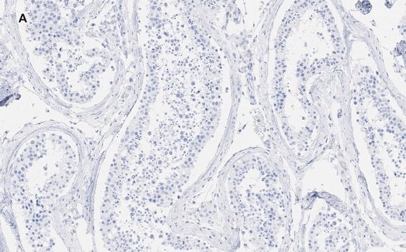
Immunocytochemistry Analysis: A 1:200 dilution from a representative lot detected YTHDF2 in HeLa cells (Chuan He, Univerity of Chicago).

Quality

Evaluated by Western Blotting in C6 cell lysate.

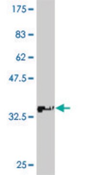
Western Blotting Analysis: 1.0 µg/mL of this antibody detected YTHDF2 in 10 µg of C6 cell lysate.

Target description

~62 kDa observed. Uncharacterized band(s) may appear in some lysates.

Physical form

Affinity purified
Purified rabbit polyclonal in buffer containing 0.1 M Tris-Glycine (pH 7.4), 150 mM NaCl with 0.05% sodium azide.

Storage and Stability

Stable for 1 year at 2-8°C from date of receipt.

Other Notes

Concentration: Please refer to lot specific datasheet.

Disclaimer

Unless otherwise stated in our catalog or other company documentation accompanying the product(s), our products are intended for research use only and are not to be used for any other purpose, which includes but is not limited to, unauthorized commercial uses, in vitro diagnostic uses, ex vivo or in vivo therapeutic uses or any type of consumption or application to humans or animals.

Not finding the right product?  

Try our Product Selector Tool.

Storage Class Code

12 - Non Combustible Liquids

WGK

WGK 1

Flash Point(F)

Not applicable

Flash Point(C)

Not applicable


Certificates of Analysis (COA)

Search for Certificates of Analysis (COA) by entering the products Lot/Batch Number. Lot and Batch Numbers can be found on a product’s label following the words ‘Lot’ or ‘Batch’.

Already Own This Product?

Find documentation for the products that you have recently purchased in the Document Library.

Visit the Document Library

Wuxun Lu et al.
The Journal of biological chemistry, 293(34), 12992-13005 (2018-07-07)
The internal N6-methyladenosine (m6A) modification of cellular mRNA regulates post-transcriptional gene expression. The YTH domain family proteins (YTHDF1-3 or Y1-3) bind to m6A-modified cellular mRNAs and modulate their metabolism and processing, thereby affecting cellular protein translation. We previously reported that
Lin Yang et al.
Communications biology, 5(1), 495-495 (2022-05-26)
The chondrogenic differentiation of bone marrow-derived mesenchymal stem cells (BMSCs) has been used in the treatment and repair of cartilage defects; however, the in-depth regulatory mechanisms by which RNA modifications are involved in this process are still poorly understood. Here
Ruifan Wu et al.
Cell death & disease, 10(3), 171-171 (2019-02-23)
Embryonic stem cells (ESCs) and induced pluripotent stem cells (iPSCs) hold great promise for regenerative medicine, disease treatment, and organ transplantation. As the ethical issue of human ESCs and similarity of pig in human genome and physiological characteristics, the porcine
Sarah N'Da Konan et al.
Retrovirology, 19(1), 4-4 (2022-02-02)
The recent discovery of the role of m6A methylation in the regulation of HIV-1 replication unveiled a novel layer of regulation for HIV gene expression. This epitranscriptomic modification of HIV-1 RNAs is under the dynamic control of specific writers and
Angelica Benavides-Serrato et al.
Cancer letters, 562, 216178-216178 (2023-04-16)
A major mechanism conferring resistance to mTOR inhibitors is activation of a salvage pathway stimulating internal ribosome entry site (IRES)-mediated mRNA translation, driving the synthesis of proteins promoting resistance of glioblastoma (GBM). Previously, we found this pathway is stimulated by

Our team of scientists has experience in all areas of research including Life Science, Material Science, Chemical Synthesis, Chromatography, Analytical and many others.

Contact Technical Service