Skip to Content
Merck
All Photos(3)

Key Documents

ABS82

Sigma-Aldrich

Anti-VEGF Antibody

from rabbit, purified by affinity chromatography

Synonym(s):

vascular endothelial growth factor A, vascular endothelial growth factor, Vascular permeability factor, vascular endothelial growth factor isoform VEGF

Sign Into View Organizational & Contract Pricing


About This Item

UNSPSC Code:
12352203
eCl@ss:
32160702
NACRES:
NA.41

biological source

rabbit

Quality Level

antibody form

affinity isolated antibody

antibody product type

primary antibodies

clone

polyclonal

purified by

affinity chromatography

species reactivity

mouse, rat, human

species reactivity (predicted by homology)

primate (based on 100% sequence homology)

technique(s)

immunohistochemistry: suitable (paraffin)
western blot: suitable

NCBI accession no.

UniProt accession no.

shipped in

wet ice

target post-translational modification

unmodified

Gene Information

human ... VEGFA(7422)

General description

VEGF (Vascular endothelial growth factor, VEGFA, VEGF-A), a dimeric ligand, is among the most potent angiogenic mitogens. VEGF is secreted by tumor cells and other cells exposed to hypoxia. VEGF is a highly specific mitogen for vascular endothelial cells. Seven VEGF isoforms (a, b, c, d, e, f, g) are generated as a result of alternative splicing from a single VEGF gene. The most commonly studied isoforms are VEGF121, VEGF165, and VEGF189 (f, d and b, respectively) All seven isoforms differ in their molecular mass and in biological properties such as their ability to bind to cell-surface heparan-sulfate proteoglycans. The expression of VEGF is potentiated and is secreted by tumor cells in response to hypoxia, by activated oncogenes, and by a variety of cytokines. VEGF induces angiogenesis as well as permeabilization of blood vessels, and plays a central role in the regulation of vasculogenesis. VEGF expression correlates to the degree of tumor vascularization and with increased metastatic risk. Additionally, VEGF induces endothelial cell proliferation, promotes cell migration, and inhibits apoptosis. Consequently, inhibition of VEGF signaling abrogates the development of a wide variety of tumors.

Specificity

Other homologies: Bovine (85% sequence homology). Rat (70% sequence homology). Mouse (65% sequence homology).
This antibody recognizes VEGF at the N-terminus.

Immunogen

Epitope: N-terminus
KLH-conjugated linear peptide corresponding to human VEGF at the N-terminus.

Application

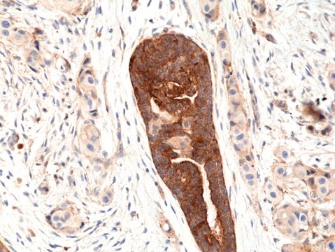
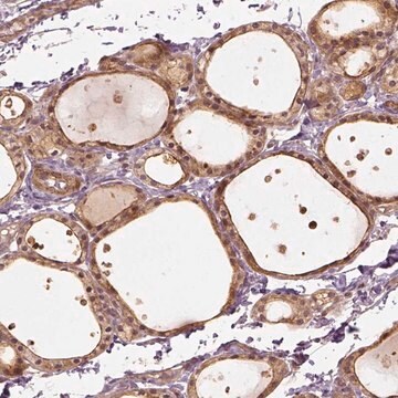
Detect VEGF using this Anti-VEGF Antibody validated for use in WB, IH(P).
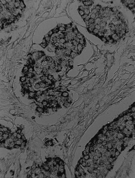
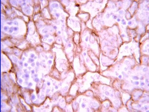
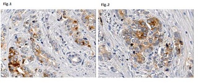
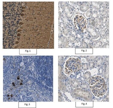
Immunohistochemistry Analysis: 1:400 dilution from a previous lot detected VEGF in normal human placental villi tissue.
Research Category
Signaling
Research Sub Category
Growth Factors & Receptors

Quality

Evaluated by Western Blot in HuVec cell lysate.

Western Blot Analysis: 0.5 µg/mL of this antibody detected VEGF on 10 µg of HuVec cell lysate.

Target description

~ 55 kDa observed. UniProtKB/Swiss-Prot P15692 (VEGFA_HUMAN) entry describes 13 isoforms produced by alternative promoter usage, alternative splicing and alternative initiation: Isoform 1 at 27.042 kDa, Isoform 2 at 25.173 kDa, Isoform 3 at 24.422 kDa, Isoform 4 at 22.314 kDa, Isoform 5 at 20.218 kDa, Isoform 6 at 20.064 kDa, Isoform 7 at 22.259 kDa, Isoform 8 at 17.219 kDa, Isoform 9 at 15.981 kDa, Isoform 10 at 40.738 kDa, Isoform 11 at 35.643 kDa, Isoform 12 at 43.597 kDa, Isoform 13 at 45.467 kDa

Linkage

Replaces: 07-1376

Physical form

Affinity purified
Purified rabbit polyclonal in buffer containing 0.1 M Tris-Glycine (pH 7.4), 150 mM NaCl with 0.05% sodium azide.

Storage and Stability

Stable for 1 year at 2-8°C from date of receipt.

Analysis Note

Control
HuVec cell lysate

Other Notes

Concentration: Please refer to the Certificate of Analysis for the lot-specific concentration.

Disclaimer

Unless otherwise stated in our catalog or other company documentation accompanying the product(s), our products are intended for research use only and are not to be used for any other purpose, which includes but is not limited to, unauthorized commercial uses, in vitro diagnostic uses, ex vivo or in vivo therapeutic uses or any type of consumption or application to humans or animals.

Not finding the right product?  

Try our Product Selector Tool.

Storage Class Code

12 - Non Combustible Liquids

WGK

WGK 1

Flash Point(F)

Not applicable

Flash Point(C)

Not applicable


Certificates of Analysis (COA)

Search for Certificates of Analysis (COA) by entering the products Lot/Batch Number. Lot and Batch Numbers can be found on a product’s label following the words ‘Lot’ or ‘Batch’.

Already Own This Product?

Find documentation for the products that you have recently purchased in the Document Library.

Visit the Document Library

Si-Yong Gao et al.
PloS one, 12(6), e0179248-e0179248 (2017-06-09)
Bisphosphonates related osteonecrosis of jaw (BRONJ) is a severe complication of systemic BPs administration, the mechanism of which is still unclarified. Recently, platelet-derived growth factor-BB (PDGF-BB) secreted by preosteoclasts was reported to promote angiogenesis and osteogenesis. This study aimed to
Airway administration of vascular endothelial growth factor siRNAs induces transient airspace enlargement in mice.
Takahashi, Y; Izumi, Y; Kohno, M; Ikeda, E; Nomori, H
International Journal of Medical Sciences null
Jiang Huang et al.
Molecular medicine reports, 12(4), 5951-5956 (2015-08-05)
Low oxygen availability is known to activate the hypoxia-inducible factor-1α (HIF-1α) pathway, which is involved in the impairment of fracture healing. However, the role of low oxygen in fracture healing remains to be fully elucidated. In the present study, rats
Chon-Kit Chou et al.
International journal of molecular sciences, 19(7) (2018-06-24)
Unfolded protein response (UPR) is a cytoprotective mechanism that alleviates the protein-folding burden in eukaryotic organisms. Moderate activation of UPR is required for maintaining endoplasmic reticulum (ER) homeostasis and profoundly contributes to tumorigenesis. Defects in UPR signaling are implicated in
Li-Fei Wang et al.
International journal of ophthalmology, 12(10), 1539-1547 (2019-10-23)
To explore the effect of Obtusifolin on retinal pigment epithelial cell growth under hypoxia. In vitro chemical hypoxia model of ARPE-19 cells was established using cobalt chloride (CoCl2). Cell viability was tested by cell counting kit-8 (CCK-8) assay. Western blot

Our team of scientists has experience in all areas of research including Life Science, Material Science, Chemical Synthesis, Chromatography, Analytical and many others.

Contact Technical Service