Skip to Content
Merck
All Photos(3)

Key Documents

MAB19562

Sigma-Aldrich

Anti-Laminin-5 (γ2 chain) Antibody, clone D4B5

clone D4B5, Chemicon®, from mouse

Synonym(s):

Laminin 5

Sign Into View Organizational & Contract Pricing


About This Item

UNSPSC Code:
12352203
eCl@ss:
32160702
NACRES:
NA.41

biological source

mouse

Quality Level

antibody form

purified immunoglobulin

antibody product type

primary antibodies

clone

D4B5, monoclonal

species reactivity

human

manufacturer/tradename

Chemicon®

technique(s)

ELISA: suitable
immunohistochemistry: suitable (paraffin)
western blot: suitable

isotype

IgG1

NCBI accession no.

UniProt accession no.

shipped in

wet ice

target post-translational modification

unmodified

Gene Information

human ... LAMC2(3918)

General description

Laminins are basement membrane, extracellular matrix glycoproteins that are composed of alpha, beta and gamma heterotrimeric chains. Different combinations of five alpha chains, three beta chains, and two gamma chains form at least 12 laminin isoforms. Laminin-5 (LN5), which consists of laminin alpha3, beta3 and gamma2 chains, is a highly conserved component of anchoring filaments underlying the hemidesmosome structure of epidermal keratinocytes. Mutations or deletion in the LN5 genes (LAMA3, LAMB3, and LAMC2) is associated with epidermolysis bullosa, a lethal skin blistering disease. LN5 may stabilize the epidermal/dermal junction of skin through binding with integrins alpha3/beta1, alpha6/beta4 and type VII collagen and has a critical role in the anchorage and locomotion of a wide variety of cell types, including epithelial cells, fibroblasts, neurons and leukocytes. Compared to fibronectin, collagen, or vitronectin, cells of epithelial origin will adhere to Laminin-5 faster and will spread to a larger extent. Recently, Laminin-5 is attracting attention as a substrate for carcinoma cells that may stimulate migration, since it has been detected at the leading edge of invasive cancer tissue in several types of human carcinomas (Niki, et al., 2002, Lohi, et al., 2001).

Specificity

Monoclonal antibody MAB19562 was developed against amino acid residues 382-608 of human laminin-5 gamma2 chain (III domain) expressed as a GST fusion protein. The antibody recognizes the 150 (precursor form) and 105 kDa (mature form) chain proteins of Laminin-5. No reactivity is expected with other laminin types, as the gamma2 chain of laminin is unique to laminin-5.

Immunogen

Amino acid residues 382-608 of human laminin-5 gamma2 chain (III domain) expressed as a GST fusion protein

Application

Anti-Laminin-5 (γ2 chain) Antibody, clone D4B5 is an antibody against Laminin-5 (γ2 chain) for use in ELISA, IH(P) & WB.
Research Category
Cell Structure
Research Sub Category
ECM Proteins
Western blot: 5ug/ml is recommended; dilute antibody in PBS or TBS only, not blocking solutions; blocking with purified compounds is suggested.

Immunohistochemistry of paraffin sections: 5-10ug/ml is suggested. Do not over fix. A light 4% or 2% PFA are best. 4 mm thick paraffin sections are dewaxed, rehydrated and immersed in 0.3% hydrogen peroxide-containing methanol for inactivation of intrinsic peroxidase, followed by treatment with Protease XXIV (Sigma) for 15 min at room temperature (0.01-0.05%). Do not use trypsin. Antibody incubation at 37°C for 1 hour, followed by secondary antibody detection and visualization with DAB (Mizushima et al. 1998). Do not place the monoclonal antibody in serum blocks because serum conatins laminin 5 which will bind and interfere with the antibody. Use BSA or other such media instead.

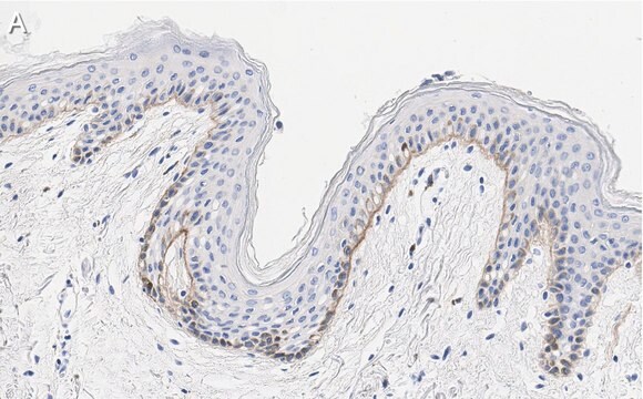
Strongly positive staining is seen in Basement Membrane of the following tissues: Skin, Esophagus, Lung, and Kidney. Positive staining is also seen in Stomach, Small intestine, Spleen, Thymus, Prostate, and Ovary. Thyroid, Testis, Skeletal and Cardiac muscle were negative for LN5 staining.

Inhibition of alpha2beta1 integrin binding to laminin 5 (40ug/ml) (Decline & Rousselle 2000).

ELISA Optimal working dilutions must be determined by end user.

Quality

Evaluated by Western Blot on recombinant Laminin-5.

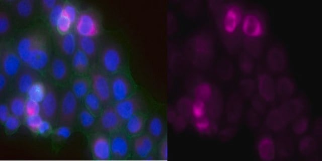
Western Blot Analysis: A 1:500-1:2000 dilution of this antibody detected Laminin-5, gamma2 chain in 10 µg of human recombinant Laminin-5.

Target description

150 kDa (precursor form) and 105 kDa (mature form)

Physical form

Format: Purified
Protein A Purified mouse immunoglobulin in 20 mM sodium phosphate, 250 mM NaCl, pH. 7.6, with 0.1% sodium azide as a preservative.
Protein A purified

Storage and Stability

Maintain for 1 year at 2–8°C from date of shipment. Aliquot to avoid repeated freezing and thawing. For maximum recovery of product, centrifuge the original vial after thawing and prior to removing the cap.

Analysis Note

Control
Human pancreatic tumor tissue, human breast carcinoma tissue or A-431 whole cell lysate

Other Notes

Concentration: Please refer to the Certificate of Analysis for the lot-specific concentration.

Legal Information

CHEMICON is a registered trademark of Merck KGaA, Darmstadt, Germany

Disclaimer

Unless otherwise stated in our catalog or other company documentation accompanying the product(s), our products are intended for research use only and are not to be used for any other purpose, which includes but is not limited to, unauthorized commercial uses, in vitro diagnostic uses, ex vivo or in vivo therapeutic uses or any type of consumption or application to humans or animals.

Not finding the right product?  

Try our Product Selector Tool.

Storage Class Code

10 - Combustible liquids

WGK

WGK 2


Certificates of Analysis (COA)

Search for Certificates of Analysis (COA) by entering the products Lot/Batch Number. Lot and Batch Numbers can be found on a product’s label following the words ‘Lot’ or ‘Batch’.

Already Own This Product?

Find documentation for the products that you have recently purchased in the Document Library.

Visit the Document Library

Kamis Gaballah et al.
Cancer research, 67(15), 7284-7294 (2007-08-03)
There is no effective medical treatment for oral precancer, and surgery to remove these lesions is imprecise because abnormal mucosa extends beyond the visible lesion. Development of vectors for tumor-selective viral replication has been a significant advance, and viral lysis
Jesica Diaz-Vera et al.
Journal of cell science, 130(4), 697-711 (2017-01-08)
We have previously shown that Rab17, a small GTPase associated with epithelial polarity, is specifically suppressed by ERK2 (also known as MAPK1) signalling to promote an invasive phenotype. However, the mechanisms through which Rab17 loss permits invasiveness, and the endosomal
Daniel Timo Behrens et al.
The Journal of biological chemistry, 287(22), 18700-18709 (2012-04-12)
The basement membrane between the epidermis and the dermis is indispensable for normal skin functions. It connects, and functionally separates, the epidermis and the dermis. To understand the suprastructural and functional basis of these connections, heterotypic supramolecular aggregates were isolated
Novel sutureless transplantation of bioadhesive-coated, freeze-dried amniotic membrane for ocular surface reconstruction.
Sekiyama, E; Nakamura, T; Kurihara, E; Cooper, LJ; Fullwood, NJ; Takaoka, M; Hamuro, J; Kinoshita, S
Investigative Ophthalmology & Visual Science null
Lionel Remy et al.
Cancer research, 66(23), 11228-11237 (2006-12-06)
Matrilysin 1 [matrix metalloproteinase 7 (MMP7)] is one of the most important metalloproteinases expressed in human tissues. This enzyme is generally not expressed by normal differentiated epithelial colon cells, but has been shown to be up-regulated in human colon adenomas

Our team of scientists has experience in all areas of research including Life Science, Material Science, Chemical Synthesis, Chromatography, Analytical and many others.

Contact Technical Service