MAB3391
Anti-Collagen Type I (COL1A1) Antibody
CHEMICON®, mouse monoclonal, 5D8-G9
Synonym(s):
Anti-CAFYD, Anti-EDSARTH1, Anti-EDSC, Anti-OI1, Anti-OI2, Anti-OI3, Anti-OI4
About This Item
Recommended Products
Product Name
Anti-Collagen Type I Antibody, clone 5D8-G9, clone 5D8-G9, Chemicon®, from mouse
biological source
mouse
Quality Level
antibody form
purified immunoglobulin
antibody product type
primary antibodies
clone
5D8-G9, monoclonal
species reactivity
pig, canine, sheep, human, bovine
should not react with
guinea pig, ground squirrel, rat, kangaroo, horse, chicken, mouse
manufacturer/tradename
Chemicon®
technique(s)
ELISA: suitable
flow cytometry: suitable
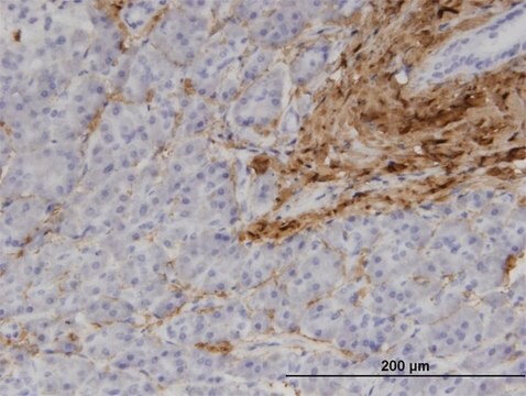
immunohistochemistry: suitable
western blot: suitable
isotype
IgG1
NCBI accession no.
UniProt accession no.
shipped in
wet ice
target post-translational modification
unmodified
Gene Information
human ... COL1A1(1277)
Related Categories
General description
Specificity
Collagen Type I Antibody displays a high affinity for human, bovine and ovine Type I Collagens. There is no evidence for cross reactivity with Collagen Types III, V and VI or connective tissue protein. Antibody reacts only with native, non-denatured Collagen I
Application
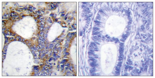
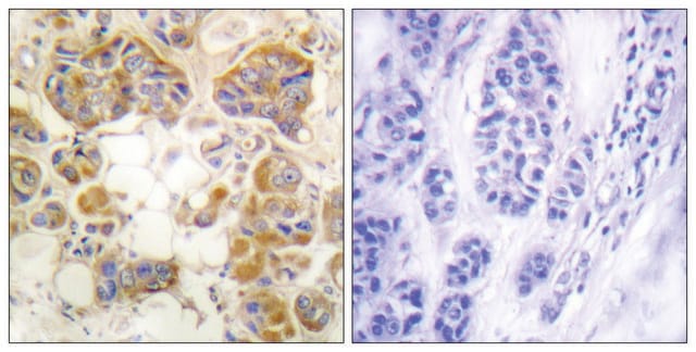
Immunohistochemistry:unfixed frozen sections:
Cut 6 μm thick sections from frozen samples using a freezing microtome. Air-dry throughly. Rehydrate with PBS. Blocking is usually not necessary for IF studies, if using DAB, pretreatment for peroxidase is suggested and blocking 2% BSA in PBS will reduce back ground.
Dilute Collagen Type I Antibody in PBS buffer, add to the sample(s) and incubate for 60 minutes. (1:100-1:500).
Wash sections for 10 minutes in PBS, twice. Add FITC conjugated anti-mouse antibody and incubate for 60 minutes.
Wash for 10 minutes in PBS, twice.
Mount sections in glycerol/water (9:1 v/v) containing 1 mM 1,4-phenylenediamine. (for FITC stability or use Chemicon catalog numbers 5096 or 5013.
NOTE:
Preliminary studies suggest that enhanced staining of certain tissues maybe obtained by pretreatment (previous to step 2) of sections with various enzymes (eg 0.1% pepsin in 0.1M HCl, 37°C for 5 minutes).
Flow Cytometry: 1:100
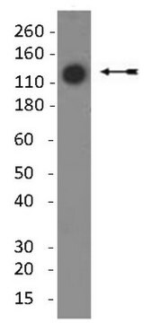
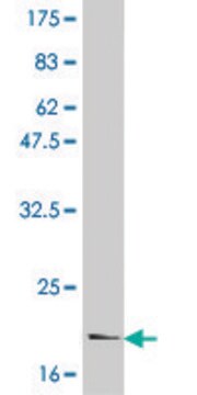
Immunoblotting:1:1000 using NATIVE, non-denatured, non-reduced western blots only. Ramshaw, et.al (1988) "Electrophoretic and electroblotting of native collagens." Anal. Biochem. 168:82-87. Antibody will not work in traditional, reduced western blots.
Briefly, PAGE gels must be prepared for running native collagens.
A 3% (w/v) total acrylamide separating gel, containing 3.2% (w/w) bis-acrylamide as a proportion to a monomer as the crosslinking agent, in 10mM calcium lactate, pH 6.8 is polymerized between vertical glass plates by the addition of 0.05-0.1% TEMED and 0.05-.1% ammonium persulfate (added from a 10% stock solution). The cathode buffer (negative) buffer is 50mM Tris adjusted to pH 6.6 with lactic acid; the anode (positive) buffer is 0.1M lactic acid pH 2.5 {Friis, SJ et al, 1985 J Biochem Biophys Methods 10:301-306.}.
Prior to sample loading the gel is run for at least 90 minutes at 100V. Collagen samples are dissolved at 1mg/mL in either 0.1M lactic acid or 0.1M acetic acid containing 10% sucrose, and 5-10μg per lane is loaded. Methyl green is added as required to assist loading. Electrophoresis is 70V for 5 hours, room temperature.
Blotting uses 0.1M lactic acid pH 2.5, and 20V for 16 hours at room temperature with the collagens migrating toward the cathode.
Cell Structure
ECM Proteins
Physical form
The immunoglobulin fraction has been purified by Protein A Chromatography and shown to be >95% pure by coomassie PAGE. The Collagen Type I Antibody is supplied 0.22 micron filtered.
Storage and Stability
WARNING: The monoclonal reagent solution contains 0.1% sodium azide as a preservative. Due to potential hazards arising from the build up of this material in pipes, spent reagent should be disposed of with liberal volumes of water.
Analysis Note
Human kidney lysate
Legal Information
Disclaimer
Not finding the right product?
Try our Product Selector Tool.
Storage Class Code
12 - Non Combustible Liquids
WGK
WGK 2
Flash Point(F)
Not applicable
Flash Point(C)
Not applicable
Certificates of Analysis (COA)
Search for Certificates of Analysis (COA) by entering the products Lot/Batch Number. Lot and Batch Numbers can be found on a product’s label following the words ‘Lot’ or ‘Batch’.
Already Own This Product?
Find documentation for the products that you have recently purchased in the Document Library.
Customers Also Viewed
Our team of scientists has experience in all areas of research including Life Science, Material Science, Chemical Synthesis, Chromatography, Analytical and many others.
Contact Technical Service