Skip to Content
Merck
All Photos(1)

Key Documents

MABN50A4

Sigma-Aldrich

Anti-Olig2 Antibody, clone 211F1.1, Alexa Fluor488 Conjugate | MABN50A4

clone 211F1.1, from mouse, ALEXA FLUOR 488

Synonym(s):

Oligodendrocyte transcription factor 2, Oligo2, Class B basic helix-loop-helix protein 1, bHLHb1, Class E basic helix-loop-helix protein 19, bHLHe19, Protein kinase C-binding protein 2, Protein kinase C-binding protein RACK17

Sign Into View Organizational & Contract Pricing


About This Item

UNSPSC Code:
12352203
eCl@ss:
32160702
NACRES:
NA.41

biological source

mouse

Quality Level

conjugate

ALEXA FLUOR 488

antibody form

purified immunoglobulin

antibody product type

primary antibodies

clone

211F1.1, monoclonal

species reactivity

rat, mouse

species reactivity (predicted by homology)

human (based on 100% sequence homology)

technique(s)

immunocytochemistry: suitable

isotype

IgG2a

NCBI accession no.

UniProt accession no.

shipped in

wet ice

target post-translational modification

unmodified

Gene Information

human ... OLIG2(10215)

General description

Oligodendrocyte lineage transcription factor 2 (Olig2) is a protein belonging to group A of the OLIG family of basic helix-loop-helix transcription factors (bHLH). bHLH proteins are divided into 6 groups (A-F), and characterized by their biochemical properties and structure. Olig2 is classified as group A due to its ability to form heterodimers with other bHLH proteins and binding to the E box. Olig2 has a crucial role during development in specifying the final location of motor neurons and oligodendrocytes in the spinal cord, along with the development within the hindbrain of somatic motor neurons. Olig2 expression is observed within oligodendrocytes and in developing astrocytes. Recent studies have shown strong expression of Olig2 in human oligodendrogliomas, primary tumors within the central nervous system, and moderate expression in astrocytomas. These observations have led to the proposal of Olig2 having potential as an oligodendroglioma diagnostic marker.

Immunogen

Recombinant protein corresponding to human Olig2.

Application

Research Category
Neuroscience
Research Sub Category
Developmental Neuroscience

Quality

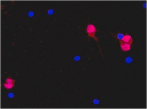
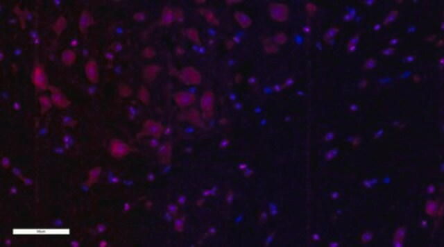
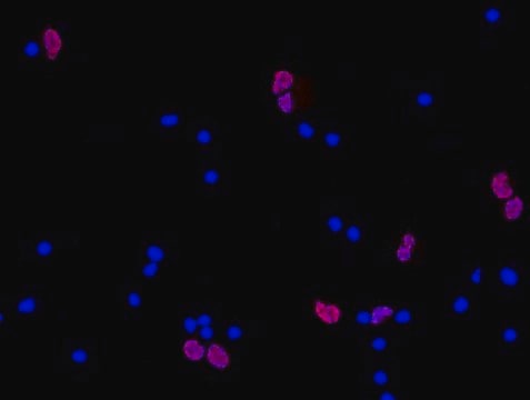
Evaluated by Immunocytochemistry in rat oligodendrocyte precursor cells.

Immunocytochemistry Analysis: A 1:100 dilution of this antibody detected Olig2 in rat oligodendrocyte precursor cells.

Target description

The unconjugated parent antibody (Cat. No. MABN50) has an observed molecular weight at ~37 kDa

Physical form

Protein G Purified
Purified mouse monoclonal IgG2a conjugated to Alexa Fluor 488 in PBS with 0.1% sodium azide and 15 mg/mL BSA.

Storage and Stability

Maintain refrigerated at 2-8 °C protected from light in undiluted aliquots for up to 6 months from date of receipt.

Analysis Note

Control
Rat oligodendrocyte precursor cells

Legal Information

ALEXA FLUOR is a trademark of Life Technologies

Disclaimer

Unless otherwise stated in our catalog or other company documentation accompanying the product(s), our products are intended for research use only and are not to be used for any other purpose, which includes but is not limited to, unauthorized commercial uses, in vitro diagnostic uses, ex vivo or in vivo therapeutic uses or any type of consumption or application to humans or animals.

Not finding the right product?  

Try our Product Selector Tool.

Storage Class Code

12 - Non Combustible Liquids

WGK

WGK 2

Flash Point(F)

Not applicable

Flash Point(C)

Not applicable


Certificates of Analysis (COA)

Search for Certificates of Analysis (COA) by entering the products Lot/Batch Number. Lot and Batch Numbers can be found on a product’s label following the words ‘Lot’ or ‘Batch’.

Already Own This Product?

Find documentation for the products that you have recently purchased in the Document Library.

Visit the Document Library

Itzy E Morales Pantoja et al.
PloS one, 15(6), e0233980-e0233980 (2020-06-09)
Multiple sclerosis (MS) is an inflammatory and demyelinating disease of the central nervous system (CNS) that results in variable severities of neurodegeneration. The understanding of MS has been limited by the inaccessibility of the affected cells and the lengthy timeframe
Sofía Puvogel et al.
Molecular psychiatry (2022-10-04)
The midbrain is an extensively studied brain region in schizophrenia, in view of its reported dopamine pathophysiology and neuroimmune changes associated with this disease. Besides the dopaminergic system, the midbrain contains other cell types that may be involved in schizophrenia
Emma Gerrits et al.
Acta neuropathologica, 141(5), 681-696 (2021-02-21)
Alzheimer's disease (AD) is the most prevalent form of dementia and is characterized by abnormal extracellular aggregates of amyloid-β and intraneuronal hyperphosphorylated tau tangles and neuropil threads. Microglia, the tissue-resident macrophages of the central nervous system (CNS), are important for
Andy P Tsai et al.
Immunity, 56(9), 2121-2136 (2023-09-03)
Genetic association studies have demonstrated the critical involvement of the microglial immune response in Alzheimer's disease (AD) pathogenesis. Phospholipase C-gamma-2 (PLCG2) is selectively expressed by microglia and functions in many immune receptor signaling pathways. In AD, PLCG2 is induced uniquely
Fumio Takahashi et al.
iScience, 25(5), 104278-104278 (2022-05-17)
Neurodegeneration is a process involving both cell autonomous and non-cell autonomous neuron loss, followed by a collapse of neural networks, but its pathogenesis is poorly understood. We have previously demonstrated that Eomes-positive helper T (Eomes + Th) cells recognizing LINE-1(L1)-derived prototypic

Our team of scientists has experience in all areas of research including Life Science, Material Science, Chemical Synthesis, Chromatography, Analytical and many others.

Contact Technical Service