Skip to Content
Merck
All Photos(1)

Key Documents

B0287

Sigma-Aldrich

Monoclonal Anti-FITC−Biotin antibody produced in mouse

clone FL-D6, purified immunoglobulin, buffered aqueous solution

Synonym(s):

Monoclonal Anti-FITC

Sign Into View Organizational & Contract Pricing


About This Item

UNSPSC Code:
12352203
NACRES:
NA.43

biological source

mouse

Quality Level

conjugate

biotin conjugate

antibody form

purified immunoglobulin

antibody product type

primary antibodies

clone

FL-D6, monoclonal

form

buffered aqueous solution

technique(s)

immunohistochemistry (formalin-fixed, paraffin-embedded sections): 1:400 using human tonsil

isotype

IgG1

shipped in

dry ice

storage temp.

−20°C

target post-translational modification

unmodified

Looking for similar products? Visit Product Comparison Guide

General description

Monoclonal Anti-FITC (mouse IgG1 isotype) is derived from the hybridoma produced by the fusion of mouse myeloma cells and splenocytes from an immunized mouse.

Specificity

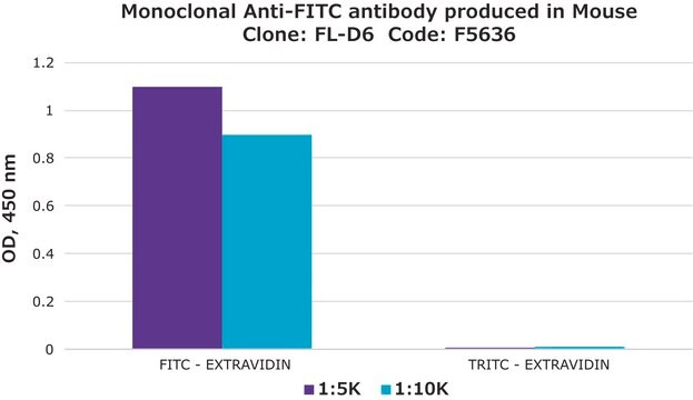
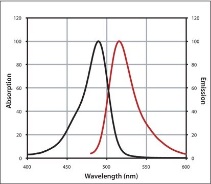
The antibody reacts with both free and conjugated fluorescein isothiocyanate (FITC).

Immunogen

FITC-BSA conjugate

Application

Monoclonal Anti-FITC-Biotin antibody produced in mouse has been used:
  • for amplification of signal in immunofluorescence assays
  • in Ag-activated beads for flow-based micro immunoassay of parathyroid hormone and IL-5
  • enzyme linked immunosorbent assay (ELISA)

Biochem/physiol Actions

Fluorochrome labeling provides a rapid and accurate localization of the site of antigen-antibody interaction when one of the reactants from parts of a cell, tissue or other biological structure. Fluorescein isothiocyanate (FITC) is a commonly used marker for antibodies in immunofluorescence techniques since the conjugation of FITC to proteins is relatively easy and does not, in general, destroy the biological activity of the labelled substances. FITC is widely used as a hapten to label different proteins and anti-FITC antibodies are used to identify these labeled proteins. Antibodies to FITC serve as universal indicator reagents by bridging FITC with another immunohistochemical reagent, such as alkaline phosphatase, horseradish peroxidase or biotin.

Physical form

Solution in 0.01 M phosphate buffered saline, pH 7.4, containing 1% bovine serum albumin, and 15 mM sodium azide

Disclaimer

Unless otherwise stated in our catalog or other company documentation accompanying the product(s), our products are intended for research use only and are not to be used for any other purpose, which includes but is not limited to, unauthorized commercial uses, in vitro diagnostic uses, ex vivo or in vivo therapeutic uses or any type of consumption or application to humans or animals.

Not finding the right product?  

Try our Product Selector Tool.

Storage Class Code

10 - Combustible liquids

WGK

WGK 3

Flash Point(F)

Not applicable

Flash Point(C)

Not applicable


Certificates of Analysis (COA)

Search for Certificates of Analysis (COA) by entering the products Lot/Batch Number. Lot and Batch Numbers can be found on a product’s label following the words ‘Lot’ or ‘Batch’.

Already Own This Product?

Find documentation for the products that you have recently purchased in the Document Library.

Visit the Document Library

Peter Stachon et al.
Arteriosclerosis, thrombosis, and vascular biology, 34(10), 2237-2245 (2014-08-12)
Nucleotides such as ATP, ADP, UTP, and UDP serve as proinflammatory danger signals via purinergic receptors on their release to the extracellular space by activated or dying cells. UDP binds to the purinergic receptor Y6 (P2Y6) and propagates vascular inflammation
Kazuyuki Kasahara et al.
Cell host & microbe, 31(6), 1038-1053 (2023-06-07)
The microbes and microbial pathways that influence host inflammatory disease progression remain largely undefined. Here, we show that variation in atherosclerosis burden is partially driven by gut microbiota and is associated with circulating levels of uric acid (UA) in mice
Chapter 11 - (Strept)avidin?Biotin Systems
Bioconjugate Techniques, 465-505 (2013)
Flow-based microimmunoassay
Hayes MA, et al.
Analytical Chemistry, 73(24), 5896-5902 (2001)
Group 2 innate lymphoid cells protect mouse heart from myocardial infarction injury via interleukin 5, eosinophils, and dendritic cells.
Liu, et al.
Cardiovascular Research, 119, 1046-1061 (2023)

Our team of scientists has experience in all areas of research including Life Science, Material Science, Chemical Synthesis, Chromatography, Analytical and many others.

Contact Technical Service