Skip to Content
Merck
All Photos(2)

Key Documents

C8114

Sigma-Aldrich

Anti-α-E-Catenin antibody produced in rabbit

IgG fraction of antiserum, buffered aqueous solution

Sign Into View Organizational & Contract Pricing


About This Item

MDL number:
UNSPSC Code:
12352203
NACRES:
NA.41
conjugate:
unconjugated
application:
ARR
ICC
WB
clone:
polyclonal
species reactivity:
rat, human, canine
citations:
12
technique(s):
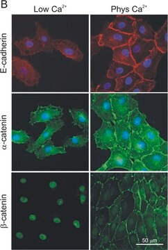
immunocytochemistry: 1:200 using methanol-fixed, dog MDCK and human MCF7 cellc
microarray: suitable
western blot: 1:1,000 using whole cell extract of human epidermal carcinoma A431 cell line and cytosolic fraction of rat embryonic brain.

biological source

rabbit

Quality Level

conjugate

unconjugated

antibody form

IgG fraction of antiserum

antibody product type

primary antibodies

clone

polyclonal

form

buffered aqueous solution

mol wt

antigen 102 kDa

species reactivity

rat, human, canine

technique(s)

immunocytochemistry: 1:200 using methanol-fixed, dog MDCK and human MCF7 cellc
microarray: suitable
western blot: 1:1,000 using whole cell extract of human epidermal carcinoma A431 cell line and cytosolic fraction of rat embryonic brain.

UniProt accession no.

shipped in

dry ice

storage temp.

−20°C

target post-translational modification

unmodified

Gene Information

General description

α-E-Catenin is a predominant subtype of α-catenin. It is widely expressed but at low levels in the nervous system. Alternative spliced variants of α-E-Catenin include α1- and α2-E-catenin.
The catenins (α, β, γ) are cytoplasmic proteins found in varying abundance in many developing and adult tissues.

Immunogen

synthetic peptide corresponding to a region located near the C-terminus of human α-E-catenin (amino acids 873-887). This sequence is identical in mouse and Xenopus α-E-catenin. It is not found in α-N-catenin, β-catenin, and γ-catenin.

Application

Anti-α-E-Catenin antibody produced in rabbit has been used in western blotting and immunocytochemistry.

Biochem/physiol Actions

Catenins bind directly or indirectly to the conserved cytoplasmic tail domain of the cell adhesion adherins. The association of catenins to cadherins produces a complex, which is linked to the actin filament network. Catenins/cadherin complexes play an important role in mediating cell adhesion, transduction of cell-cell contact positional signals to the cell interior, and may play a crucial role in cell differentiation. The linkage of the epithelial cadherin /uvomorulin to actin is essential for the cell binding function of this cadherin. α-Catenin (CAP102, 102 kDa), originally described as an E-cadherin associated protein, has been shown to associate with other members of the cadherin family members, N-cadherin and P-cadherin. Within its conserved region α-catenin shows 30% identity to vinculin.

Physical form

Solution in 0.01 M phosphate buffered saline, pH 7.4, containing 15 mM sodium azide.

Disclaimer

Unless otherwise stated in our catalog or other company documentation accompanying the product(s), our products are intended for research use only and are not to be used for any other purpose, which includes but is not limited to, unauthorized commercial uses, in vitro diagnostic uses, ex vivo or in vivo therapeutic uses or any type of consumption or application to humans or animals.

Not finding the right product?  

Try our Product Selector Tool.

Storage Class Code

10 - Combustible liquids

WGK

WGK 2

Flash Point(F)

Not applicable

Flash Point(C)

Not applicable

Personal Protective Equipment

dust mask type N95 (US), Eyeshields, Gloves

Choose from one of the most recent versions:

Certificates of Analysis (COA)

Lot/Batch Number

Don't see the Right Version?

If you require a particular version, you can look up a specific certificate by the Lot or Batch number.

Already Own This Product?

Find documentation for the products that you have recently purchased in the Document Library.

Visit the Document Library

Evolving form to fit function: cardiomyocyte intercalated disc and transverse-tubule membranes
Current Topics in Membranes, 72, 121-158 (2013)
Arad Soffer et al.
PLoS biology, 20(8), e3001756-e3001756 (2022-08-16)
Mitotic spindle orientation (SO) is a conserved mechanism that governs cell fate and tissue morphogenesis. In the developing epidermis, a balance between self-renewing symmetric divisions and differentiative asymmetric divisions is necessary for normal development. While the cellular machinery that executes
p120 catenin is required for normal renal tubulogenesis and glomerulogenesis
Marciano DK, et al.
Development, 138(10), 2099-2109 (2011)
Adnan Mahly et al.
BMC biology, 20(1), 145-145 (2022-06-17)
The establishment of tissue architecture requires coordination between distinct processes including basement membrane assembly, cell adhesion, and polarity; however, the underlying mechanisms remain poorly understood. The actin cytoskeleton is ideally situated to orchestrate tissue morphogenesis due to its roles in
A Nagafuchi et al.
Cell regulation, 1(1), 37-44 (1989-11-01)
Cadherins are a family of transmembrane glycoproteins which play a key role in Ca(2+)-dependent cell-cell adhesion. Cytoplasmic domains of these molecules are anchored to the cell cytoskeleton and are required for cadherin function. To elucidate how the function of cadherins

Our team of scientists has experience in all areas of research including Life Science, Material Science, Chemical Synthesis, Chromatography, Analytical and many others.

Contact Technical Service