Skip to Content
Merck
All Photos(1)

Key Documents

G4546

Sigma-Aldrich

Anti-Glial Fibrillary Acidic Protein Antibody

rabbit polyclonal

Synonym(s):

GFAP Antibody for Flow Cytometry - Anti-Glial Fibrillary Acidic Protein antibody produced in rabbit, Gfap Antibody Flow Cytometry, Anti-FLJ45472, Anti-GFAP, Anti-Intermediate filament protein

Sign Into View Organizational & Contract Pricing


About This Item

MDL number:
UNSPSC Code:
12352203
NACRES:
NA.41

Product Name

Anti-Glial Fibrillary Acidic Protein antibody produced in rabbit, ~1 mg/mL, affinity isolated antibody, buffered aqueous solution

biological source

rabbit

Quality Level

conjugate

unconjugated

antibody form

affinity isolated antibody

antibody product type

primary antibodies

clone

polyclonal

form

buffered aqueous solution

mol wt

antigen ~50 kDa

species reactivity

human, rat, mouse

concentration

~1 mg/mL

technique(s)

flow cytometry: 1:200-1:500
immunohistochemistry: 1:100-1:200
western blot: 1:500-1:1,000

UniProt accession no.

shipped in

dry ice

storage temp.

−20°C

target post-translational modification

unmodified

Gene Information

human ... GFAP(2670)
mouse ... Gfap(14580)
rat ... Gfap(24387)

General description

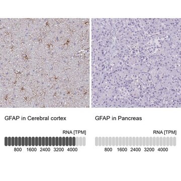
Intermediate filaments (IFs) with characteristic 10 nm diameter are a distinct class of molecularly heterogenous cytoskeletal filaments defined by ultrastructural, immunological, and biochemical criteria. Intermediate filaments differ significantly from the other cytoskeletal elements of the cell, namely microtubules and microfilaments, and are components of most eukaryotic cells. Glial Fibrillary Acidic Protein (GFAP) (molecular weight of 50 kDa) is found in astrocytes and ependymal cells of the central nervous system. It is found in various other cell types including stellate cells, chondrocytes, osteocytes, keratinocytes, Leydig cells and fibroblasts. GFAP is involved in mitosis, and cell:cell interactions and communications. Mutations in this gene cause Alexander disease, a rare disorder of astrocytes in the central nervous system.

Specificity

Rabbit polyclonal anti-Glial Fibrillary Acidic Protein recognizes human, mouse, and rat GFAPs.

Immunogen

synthetic peptide corresponding to an internal C-terminal sequence of human GFAP.

Application

Rabbit polyclonal anti-GFAP antibody is used to tag glial fibrillary acidic protein for detection and quantitation by Western blotting and immunohistochemical (IHC) techniques. It is used as a probe to determine the roles of glial fibrillary acidic protein in cell:cell communications, mitosis and cell structure. Anti-GFAP has proven a valuable tool for use in immunocytochemical localization of GFAP in normal central nervous system tissue, certain tumors and metastases of the glial antigen, as well as for immunofluorescent labeling of cultured mammalian cells.

Physical form

solution in phosphate buffered saline, containing 0.02% sodium azide.

Disclaimer

Unless otherwise stated in our catalog or other company documentation accompanying the product(s), our products are intended for research use only and are not to be used for any other purpose, which includes but is not limited to, unauthorized commercial uses, in vitro diagnostic uses, ex vivo or in vivo therapeutic uses or any type of consumption or application to humans or animals.

Not finding the right product?  

Try our Product Selector Tool.

Storage Class Code

12 - Non Combustible Liquids

WGK

nwg

Flash Point(F)

Not applicable

Flash Point(C)

Not applicable

Personal Protective Equipment

dust mask type N95 (US), Eyeshields, Gloves

Choose from one of the most recent versions:

Certificates of Analysis (COA)

Lot/Batch Number

Don't see the Right Version?

If you require a particular version, you can look up a specific certificate by the Lot or Batch number.

Already Own This Product?

Find documentation for the products that you have recently purchased in the Document Library.

Visit the Document Library

Jesús Agulla et al.
Theranostics, 4(1), 90-105 (2014-01-08)
The use of theranostics in neurosciences has been rare to date because of the limitations imposed on the free delivery of substances to the brain by the blood-brain barrier. Here we report the development of a theranostic system for the
Christopher G Dengler et al.
Scientific reports, 7, 42090-42090 (2017-02-22)
In a mouse model of temporal lobe epilepsy, multicellular calcium imaging revealed that disease emergence was accompanied by massive amplification in the normally sparse, afferent stimulation-induced activation of hippocampal dentate granule cells. Patch recordings demonstrated reductions in local inhibitory function
Arpita Konar et al.
Evidence-based complementary and alternative medicine : eCAM, 2015, 837012-837012 (2015-09-29)
Preclinical studies on animal models have discerned the antiamnesic and memory-enhancing potential of Bacopa monniera (Brahmi) crude extract and standardized extracts. These studies primarily focus on behavioral consequences. However, lack of information on molecular underpinnings has limited the clinical trials
Yanli Zhu et al.
Molecular vision, 19, 2360-2372 (2013-12-10)
We recently demonstrated in a mouse model of glaucoma that endogenous epigenetic mechanisms can be activated by a repetitive hypoxic preconditioning (RHP) stimulus to provide robust retinal ganglion cell (RGC) protection. Although we also provided evidence that RHP prevents or
Smriti M Agrawal et al.
Brain : a journal of neurology, 136(Pt 6), 1760-1777 (2013-05-21)
Inflammatory perivascular cuffs are comprised of leucocytes that accumulate in the perivascular space around post-capillary venules before their infiltration into the parenchyma of the central nervous system. Inflammatory perivascular cuffs are commonly found in the central nervous system of patients

Our team of scientists has experience in all areas of research including Life Science, Material Science, Chemical Synthesis, Chromatography, Analytical and many others.

Contact Technical Service