Skip to Content
Merck
All Photos(2)

Key Documents

M3567

Sigma-Aldrich

Anti-Myosin Iβ (Nuclear) antibody produced in rabbit

affinity isolated antibody, buffered aqueous solution

Synonym(s):

Anti-Myh 2, Anti-Myo1c, Anti-Myr 2

Sign Into View Organizational & Contract Pricing


About This Item

MDL number:
UNSPSC Code:
12352203
NACRES:
NA.41

biological source

rabbit

Quality Level

conjugate

unconjugated

antibody form

affinity isolated antibody

antibody product type

primary antibodies

clone

polyclonal

form

buffered aqueous solution

mol wt

antigen ~120 kDa

species reactivity

human, mouse, canine, rat

technique(s)

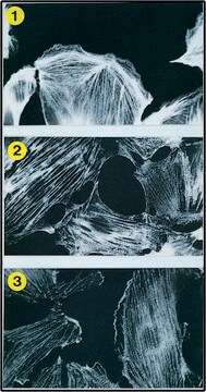
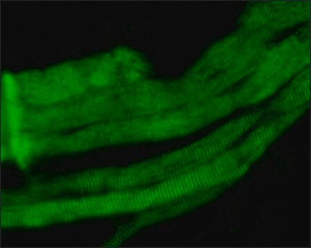
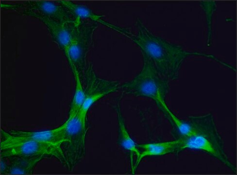
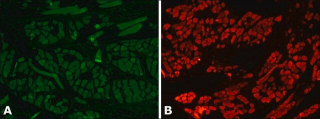
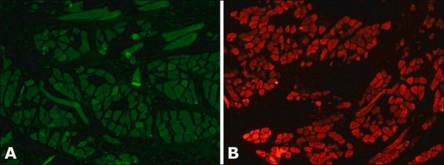
indirect immunofluorescence: 1:50 using mouse NIH3T3 cells
microarray: suitable
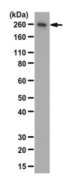
western blot: 1:1,000 using extract of human HeLa cells nuclei and dog MDCK cells

UniProt accession no.

shipped in

dry ice

storage temp.

−20°C

target post-translational modification

unmodified

Gene Information

human ... MYO1C(4641)
mouse ... Myo1c(17913)
rat ... Myo1c(65261)

General description

Myosins belong to a superfamily of actin-based motor proteins comprising to date at least 15 classes. Myosin I (also designated myosin-1c, myo1c, M1C, myr 2 and myh 2) is a widely distributed single-headed myosin composed of an N-terminal motor domain, a calmodulin-light chain binding neck region, and a short C-terminal domain. In mammalian cells, myosin I is usually found in the cytoplasm and is especially enriched in perinuclear regions and dynamic cell margins. Myosin Iβ is found in a variety of cells such as kidney tubular cells and inner-ear hair cells.

Specificity

Recognizes the heavy chain of the nuclear form of human myosin Iβ. Additional bands may be detected in some preparations.

Immunogen

synthetic peptide corresponding to amino acid residues 1-16 of the heavy chain of mouse myosin Iβ (nuclear form). This sequence is missing from the other myosin I isoforms as well as from other currently known myosins.

Application

Anti-Myosin Iβ (Nuclear) antibody produced in rabbit has been used in:
  • western blot
  • confocal immunofluorescence
  • immunogold labeling
  • immunofluorescence
  • co-immunoprecipitation experiments
  • immunostaining

Biochem/physiol Actions

Myosin I is involved in cell motility, microvilli anchorage, vesicular and organelle transport and signal transduction. Myosin I associate with lipid membranes through its tail domain while its motor domain interacts with actin filaments. Myosin Iβ (nuclear) co-localizes with RNA polymerases I and II and possibly acts as a molecular motor to power transcription.

Physical form

Solution in 0.01 M phosphate buffered saline, pH 7.4, containing 1% bovine serum albumin and 15 mM sodium azide.

Disclaimer

Unless otherwise stated in our catalog or other company documentation accompanying the product(s), our products are intended for research use only and are not to be used for any other purpose, which includes but is not limited to, unauthorized commercial uses, in vitro diagnostic uses, ex vivo or in vivo therapeutic uses or any type of consumption or application to humans or animals.

Not finding the right product?  

Try our Product Selector Tool.

Storage Class Code

10 - Combustible liquids

WGK

WGK 3

Flash Point(F)

Not applicable

Flash Point(C)

Not applicable


Choose from one of the most recent versions:

Certificates of Analysis (COA)

Lot/Batch Number

Don't see the Right Version?

If you require a particular version, you can look up a specific certificate by the Lot or Batch number.

Already Own This Product?

Find documentation for the products that you have recently purchased in the Document Library.

Visit the Document Library

Motor domain-dependent localization of myo1b (myr-1)
Tang N, et al.
Current Biology, 11(14), 1131-1135 (2001)
Subnuclear compartmentalization and function of actin and nuclear Myosin I in plants
Cruz JR, et al.
Chromosoma, 118(2), 193-207 (2009)
Nuclear myosin I regulates cell membrane tension
Venit T, et al.
Scientific Reports, 30864-30864 (2016)
Specific nuclear localizing sequence directs two myosin isoforms to the cell nucleus in calmodulin-sensitive manner
Dzijak R, et al.
PLoS ONE, 7(1), e30529-e30529 (2012)
Force Generation by Membrane-Associated Myosin-I
Pyrpassopoulos S, et al.
Scientific reports (2016)

Our team of scientists has experience in all areas of research including Life Science, Material Science, Chemical Synthesis, Chromatography, Analytical and many others.

Contact Technical Service