N6655
Anti-Nerve Growth Factor 2.5S antibody produced in rabbit
fractionated antiserum, buffered aqueous solution
Synonym(s):
Anti-NGF-2.5S
Sign Into View Organizational & Contract Pricing
All Photos(1)
About This Item
clone:
polyclonal
application:
ODD
WB
WB
species reactivity:
mouse, human
technique(s):
Ouchterlony double diffusion: suitable
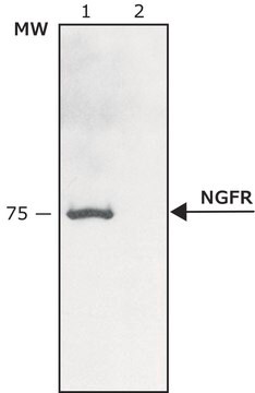
western blot: 1:2,000
western blot: 1:2,000
citations:
17
Recommended Products
biological source
rabbit
Quality Level
conjugate
unconjugated
antibody form
fractionated antiserum
antibody product type
primary antibodies
clone
polyclonal
form
buffered aqueous solution
species reactivity
mouse, human
technique(s)
Ouchterlony double diffusion: suitable
western blot: 1:2,000
UniProt accession no.
shipped in
dry ice
storage temp.
−20°C
target post-translational modification
unmodified
Gene Information
human ... NGF(4803)
mouse ... Ngf(18049)
General description
Nerve growth factor (NGF) belongs to the family of neurotrophic factors. It is located at 1p13.2 on the human chromosome. There are two molecular forms of NGF, 7S NGF and 2.5S NGF, that have identical neurotrophic effect on neurons.
Specificity
May be used in monitoring for NGF (2.5S, β and 7S).
Immunogen
NGF-2.5S from male mouse submaxillary glands.
Application
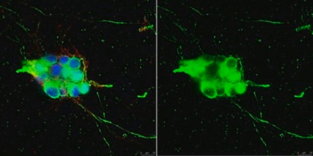
Anti-Nerve Growth Factor 2.5S antibody may be used for immunoblotting and dot blot at a minimum antibody working dilution of 1:2000. Antibody dilution of 1:1000 was used for immunoblotting using extracts of nucleus raphe magus tissue of rats. The antibody was used for immunofluorescence of mouse astrocytes and for neutralization assays.
Applications in which this antibody has been used successfully, and the associated peer-reviewed papers, are given below.
Immunofluorescence (1 paper)
Western Blotting (1 paper)
Immunofluorescence (1 paper)
Western Blotting (1 paper)
Biochem/physiol Actions
Mutations in this gene leads to hereditary sensory and autonomic neuropathies class five (HSAN-V).
Physical form
Solution in phosphate buffered saline.
Disclaimer
Unless otherwise stated in our catalog or other company documentation accompanying the product(s), our products are intended for research use only and are not to be used for any other purpose, which includes but is not limited to, unauthorized commercial uses, in vitro diagnostic uses, ex vivo or in vivo therapeutic uses or any type of consumption or application to humans or animals.
Not finding the right product?
Try our Product Selector Tool.
Storage Class Code
10 - Combustible liquids
WGK
WGK 3
Flash Point(F)
Not applicable
Flash Point(C)
Not applicable
Choose from one of the most recent versions:
Already Own This Product?
Find documentation for the products that you have recently purchased in the Document Library.
Customers Also Viewed
New protein fold revealed by a 2.3-
McDonald N Q, et al.
Nature, 354(6352), 411-411 (1991)
Liang-Wei Chen et al.
The Journal of comparative neurology, 497(6), 898-909 (2006-06-28)
To address the hypothesis that reactive astrocytes in the basal ganglia of an animal model of Parkinson's disease serve neurotrophic roles, we studied the expression pattern of neurotrophic factors in the basal ganglia of C57/Bl mice that had been treated
7S nerve growth factor has different biological activity from 2.5 S nerve growth factor in vitro
Shao N, et al.
Brain Research, 609(1-2), 338-340 (1993)
Micaela Caserta et al.
Frontiers in neuroscience, 13, 58-58 (2019-02-23)
Previous studies demonstrated exercise-induced modulation of neurotrophins, such as Nerve Growth Factor (NGF) and Brain-Derived Neurotrophic Factor (BDNF). Yet, no study that we are aware of has examined their change as a function of different training paradigms. In addition, the
Bihua Bie et al.
The Journal of neuroscience : the official journal of the Society for Neuroscience, 30(16), 5617-5628 (2010-04-23)
Sorting of intracellular G-protein-coupled receptors (GPCRs) either to lysosomes for degradation or to plasma membrane for surface insertion and functional expression is a key process regulating signaling strength of GPCRs across the plasma membrane in adult mammalian cells. However, little
Our team of scientists has experience in all areas of research including Life Science, Material Science, Chemical Synthesis, Chromatography, Analytical and many others.
Contact Technical Service