Skip to Content
Merck
All Photos(1)

Documents

PKH26PCL

Sigma-Aldrich

PKH26 Red Fluorescent Cell Linker Kit for Phagocytic Cell Labeling

Distributed for Phanos Technologies

Synonym(s):

Phagocytic cell label

Sign Into View Organizational & Contract Pricing


About This Item

UNSPSC Code:
12352207
NACRES:
NA.32

Application

This kit is for phagocytic cell labeling. It is used to selectively label cells with phagocytic capabilities such as monocytes, macrophages or neutrophils.

Principle

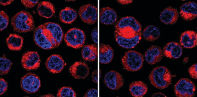
The labeling occurs through the formation of dye aggregates or particulates. The aggregate formation significantly inhibits the uptake of dye by non-phagocytic cells, such as lymphocytes, but facilitates dye uptake by phagocytic cells. Labeled cells appear patchy or spotted because the dye is localized in phagocytic compartments of the cells. The dye appears to be resistant to metabolic attack and has been found to remain with the cells for at least 21 days in vivo.
Labeling of phagocytic cells by this methodology may be conducted either in vitro or in vivo. Intraperitoneal or intravenous injections of the PKH26 labeling solution will successfully label phagocytic cells in vivo, while cells of interest which have been isolated may be stained using in vitro labeling methods.

Linkage

For additional technical details on PKH and CellVue® Fluorescent Cell Linker Dyes including an extensive bibliography, please visit here.

Legal Information

CellVue is a registered trademark of Phanos Technologies

Kit Components Only

Product No.
Description

  • Diluent B 6 x 10

  • PKH26 cell linker in ethanol .5 mL

related product

Pictograms

FlameExclamation mark

Signal Word

Danger

Hazard Statements

Hazard Classifications

Eye Irrit. 2 - Flam. Liq. 2

Storage Class Code

3 - Flammable liquids

Flash Point(F)

57.2 °F - closed cup

Flash Point(C)

14.0 °C - closed cup


Certificates of Analysis (COA)

Search for Certificates of Analysis (COA) by entering the products Lot/Batch Number. Lot and Batch Numbers can be found on a product’s label following the words ‘Lot’ or ‘Batch’.

Already Own This Product?

Find documentation for the products that you have recently purchased in the Document Library.

Visit the Document Library

K Tabata et al.
Gene therapy, 18(10), 969-978 (2011-04-23)
We previously identified the mouse and human Glipr1 and GLIPR1/RTVP-1 genes, respectively, as direct p53 targets with proapoptotic activities in various cancer cell lines, including prostate cancer (PCa). Intratumoral injection of an adenoviral vector capable of efficient transduction and expression
Jeffrey H Jennings et al.
American journal of respiratory cell and molecular biology, 32(2), 108-117 (2004-11-26)
Apoptotic cells must be cleared to resolve inflammation, but few resident alveolar macrophages (AMo) from normal lungs ingest apoptotic cells. We examined how Mo ingestion of apoptotic cells is altered during immune inflammation induced by intratracheal challenge of primed C57BL/6
Hansang Cho et al.
Lab on a chip, 14(5), 972-978 (2014-01-17)
Neutrophils are the most abundant type of white blood cells in the circulation, protecting the body against pathogens and responding early to inflammation. Although we understand how neutrophils respond to individual stimuli, we know less about how they prioritize between
Hiroshi Kanno et al.
International journal of molecular sciences, 14(5), 9604-9617 (2013-05-07)
A specialized population of cells residing in the hair follicle is quiescent but shows pluripotency for differentiating into epithelial-mesenchymal lineage cells. Therefore, such cells are hoped to be useful as implantable donor cells for regenerative therapy. Recently, it was reported
Sung Hoon Baik et al.
Neurobiology of aging, 35(6), 1286-1292 (2014-02-04)
Immune responses in the brain are thought to play a role in disorders of the central nervous system, but an understanding of the process underlying how immune cells get into the brain and their fate there remains unclear. In this

Articles

PKH dyes are easy to use and achieve stable, uniform, and reproducible fluorescent labeling of live cells. PKH dyes are non-toxic membrane stains which produce high signal to noise ratio.

Lipophilic cell tracking dyes enable cancer biologists to track tumor and immune cell functions both in vitro and in vivo. Read the article to choose a right membrane dye kit for cell tracking and proliferation monitoring.

Optimal staining is a key component for studying tumorigenesis and progression. Learn useful tips and techniques for dye applications, including examples from recent studies.

PKH and CellVue® Fluorescent Cell Linker Kits provide fluorescent labeling of live cells over an extended period of time, with no apparent toxic effects.

Our team of scientists has experience in all areas of research including Life Science, Material Science, Chemical Synthesis, Chromatography, Analytical and many others.

Contact Technical Service