Skip to Content
Merck
All Photos(1)

Key Documents

07-018-I

Sigma-Aldrich

Anti-phospho-p70 S6 Kinase (Thr389) Antibody

from rabbit, purified by affinity chromatography

Synonym(s):

Ribosomal protein S6 kinase, S6K-beta-1, S6K1, p70S6K1, p70 S6K1, p70-S6K1, p70 S6KA

Sign Into View Organizational & Contract Pricing


About This Item

UNSPSC Code:
12352203
eCl@ss:
32160702
NACRES:
NA.41
clone:
polyclonal
application:
WB
inhibition assay
species reactivity:
human
technique(s):
inhibition assay: suitable (peptide)
western blot: suitable
citations:
7

biological source

rabbit

Quality Level

antibody form

affinity isolated antibody

antibody product type

primary antibodies

clone

polyclonal

purified by

affinity chromatography

species reactivity

human

species reactivity (predicted by homology)

zebrafish (based on 100% sequence homology), chicken (based on 100% sequence homology), rat (based on 100% sequence homology), bovine (based on 100% sequence homology), mouse (based on 100% sequence homology)

technique(s)

inhibition assay: suitable (peptide)
western blot: suitable

NCBI accession no.

UniProt accession no.

shipped in

wet ice

target post-translational modification

phosphorylation (pThr389)

Gene Information

human ... RPS6KB1(6198)

General description

The p70 S6 kinase is a ubiquitous cytoplasmic protein that is activated in response to cytokines, and growth factors including the epidermal growth factor and fibroblast growth factor. It lies downstream of the mTOR/PI3K pathway and is phosphorylated on multiple residues: Thr389, Thr229, Thr421, Ser371, Ser404, Ser411, Ser418, and Ser424. Thr389, Ser371, and Ser404 are located in the linker domain; whereas Thr421, Ser411, Ser418, Ser424 are located in the autoinhibitory domain, and Thr229 is found in the catalytic domain. These sites may be phosphorylated in a hierarchical sequence leading to the activation of the p70 S6 kinase. The p70 S6 kinase phosphorylates the S6 protein on the 40S ribosomal protein (rpS6), and plays a role in multiple processes including regulation of mRNA molecules with 5’-terminal oligopyrimidine tracts, protein synthesis, cell growth, proliferation, and cell survival. The activity of this kinase may be important in diabetes, cancer, and aging.

Specificity

Other homologies: ribosomal protein S6 kinase beta-2 (91% sequence homology).
This antibody recognizes the AGC-kinase C-terminal of Ribosomal protein S6 kinase beta-1 phosphorylated at Thr389.

Immunogen

Epitope: AGC-kinase C-terminal
KLH-conjugated linear peptide corresponding to the AGC-kinase C-terminal of human Ribosomal protein S6 kinase beta-1
phosphorylated at Thr389.

Application

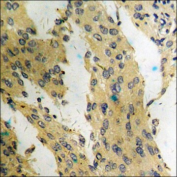
Detect phospho-p70 S6 Kinase (Thr389) using this Anti-phospho-p70 S6 Kinase (Thr389) Antibody validated for use in Western Blotting, Peptide Inhibition Assay.
Research Category
Signaling
Research Sub Category
Kinases & Phosphatases

Quality

Evaluated by Western Blot (Peptide Inhibition) Analysis in MCF7 cell lysate and MCF7 cell lysate with non-phospho or phospho peptide.

Western Blot (Peptide Inhibition) Analysis: 1.0 µg/mL of this antibody detected p70 S6 Kinase phosphorylated at Thr389 in 10 µg of MCF7 cell lysate and MCF7 cell lysate incubated with a non-phospho peptide.

Target description

~70 kDa observed

Physical form

Affinity purified
Purified rabbit polyclonal in buffer containing 0.1 M Tris-Glycine (pH 7.4), 150 mM NaCl with 0.05% sodium azide.

Storage and Stability

Stable for 1 year at 2-8°C from date of receipt.

Analysis Note

Control
MCF7 cell lysate

Other Notes

Concentration: Please refer to the Certificate of Analysis for the lot-specific concentration.

Disclaimer

Unless otherwise stated in our catalog or other company documentation accompanying the product(s), our products are intended for research use only and are not to be used for any other purpose, which includes but is not limited to, unauthorized commercial uses, in vitro diagnostic uses, ex vivo or in vivo therapeutic uses or any type of consumption or application to humans or animals.

Not finding the right product?  

Try our Product Selector Tool.

Storage Class Code

12 - Non Combustible Liquids

WGK

WGK 1

Flash Point(F)

Not applicable

Flash Point(C)

Not applicable


Certificates of Analysis (COA)

Search for Certificates of Analysis (COA) by entering the products Lot/Batch Number. Lot and Batch Numbers can be found on a product’s label following the words ‘Lot’ or ‘Batch’.

Already Own This Product?

Find documentation for the products that you have recently purchased in the Document Library.

Visit the Document Library

Iryna Stryapunina et al.
PLoS genetics, 20(1), e1011145-e1011145 (2024-01-29)
Females from many mosquito species feed on blood to acquire nutrients for egg development. The oogenetic cycle has been characterized in the arboviral vector Aedes aegypti, where after a bloodmeal, the lipid transporter lipophorin (Lp) shuttles lipids from the midgut
Nathan M Belliveau et al.
Nature communications, 14(1), 5770-5770 (2023-09-19)
Neutrophils are the most abundant leukocyte in humans and provide a critical early line of defense as part of our innate immune system. We perform a comprehensive, genome-wide assessment of the molecular factors critical to proliferation, differentiation, and cell migration
Samer Wassim El-Kadi et al.
Journal of applied physiology (Bethesda, Md. : 1985) (2018-08-03)
Muscle hypertrophy is limited in low-birth-weight (LBWT) neonates, suggesting a reduction in protein synthesis and increased protein degradation. Sixteen pairs of 1-d old normal-birth-weight (NBWT) and LBWT littermates (n = 16) were euthanized and the longissimus dorsi (LD) was sampled
Ying Chen et al.
Frontiers in physiology, 8, 482-482 (2017-07-27)
Low-birth-weight (LBWT) neonates experience restricted muscle growth in their perinatal life. Our aim was to investigate the mechanisms that contribute to slower skeletal muscle growth of LBWT neonatal pigs. Twenty-four 1-day old male LBWT (816 ± 55 g) and normal-birth-weight
A De Leo et al.
Cell death & disease, 6, e1876-e1876 (2015-09-04)
Autophagy, an important degradation system involved in maintaining cellular homeostasis, serves also to eliminate pathogens and process their fragments for presentation to the immune system. Several viruses have been shown to interact with the host autophagic machinery to suppress or

Our team of scientists has experience in all areas of research including Life Science, Material Science, Chemical Synthesis, Chromatography, Analytical and many others.

Contact Technical Service