Skip to Content
Merck
All Photos(3)

Key Documents

AB2219

Sigma-Aldrich

Anti-Aquaporin 1 Antibody

1.0 mg/mL, Chemicon®

Synonym(s):

Aquaporin-1, AQP-1, Aquaporin-CHIP, Water channel protein for red blood cells and kidney proximal tubule

Sign Into View Organizational & Contract Pricing


About This Item

UNSPSC Code:
12352203
eCl@ss:
32160702
NACRES:
NA.41
clone:
polyclonal
application:
IHC
WB
species reactivity:
rat, mouse, human
technique(s):
immunohistochemistry: suitable
western blot: suitable
citations:
64

biological source

rabbit

Quality Level

antibody form

affinity purified immunoglobulin

antibody product type

primary antibodies

clone

polyclonal

purified by

affinity chromatography

species reactivity

rat, mouse, human

species reactivity (predicted by homology)

sheep (based on 100% sequence homology), bovine (based on 100% sequence homology), primate (based on 100% sequence homology)

manufacturer/tradename

Chemicon®

concentration

1.0 mg/mL

technique(s)

immunohistochemistry: suitable
western blot: suitable

NCBI accession no.

UniProt accession no.

shipped in

wet ice

target post-translational modification

unmodified

Gene Information

human ... AQP1(358)

General description

Aquaporin 1 is an integral membrane protein which was originally identified in red blood cells and renal proximal tubules. AQP1 is also expressed by the choroid plexus and various other tissues. It forms a water-specific channel that provides the plasma membranes of red cells and kidney proximal tubules with high permeability to water, thereby permitting water to move in the direction of an osmotic gradient.

Immunogen

Epitope: Topological domain
KLH-conjugated linear peptide corresponding to the topological domain of rat Aquaporin 1.

Application

Anti-Aquaporin 1 Antibody detects level of Aquaporin 1 & has been published & validated for use in ELISA, WB, IH.
Research Category
Neuroscience
Research Sub Category
Developmental Signaling
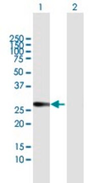
Western Blotting: 1-10 μg/mL using Chemiluminescence technique. Immunoblots reveal two bands, a lower 28 kDa band corresponding to unglycosylated form and an upper high molecular 35-60 kDa glycosylated component (Denker et al. 1988; Smith & Agre 1991) We recommend the use of 0.5-1.0% milk in all primary/secondary antibody-enzyme conjugate incubations in order to suppress non-specific bands.
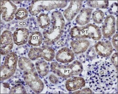
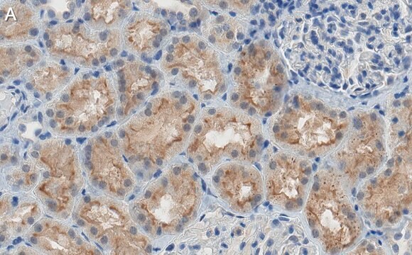
Immunohistochemistry Analysis: A 1:2,000 dilution from a representative lot detected Aquaporin 1 in human kidney and mouse kidney tissue.

Quality

Evaluated by Western Blotting in human kidney tissue lysate.

Western Blotting Analysis: 2.0 µg/mL of this antibody detected Aquaporin 1 in 10 µg of human kidney tissue lysate.

Target description

28 kDa

Linkage

Replaces: AB3065

Physical form

ImmunoAffinity Purified
Purified rabbit polyclonal in buffer containing 0.1 M Tris-Glycine (pH 7.4), 150 mM NaCl with 0.05% sodium azide.

Storage and Stability

Maintain at 2-8°C in undiluted aliquots for up to 1 year after date of receipt.

Legal Information

CHEMICON is a registered trademark of Merck KGaA, Darmstadt, Germany

Disclaimer

Unless otherwise stated in our catalog or other company documentation accompanying the product(s), our products are intended for research use only and are not to be used for any other purpose, which includes but is not limited to, unauthorized commercial uses, in vitro diagnostic uses, ex vivo or in vivo therapeutic uses or any type of consumption or application to humans or animals.

Not finding the right product?  

Try our Product Selector Tool.

Storage Class Code

12 - Non Combustible Liquids

WGK

WGK 1

Flash Point(F)

Not applicable

Flash Point(C)

Not applicable


Certificates of Analysis (COA)

Search for Certificates of Analysis (COA) by entering the products Lot/Batch Number. Lot and Batch Numbers can be found on a product’s label following the words ‘Lot’ or ‘Batch’.

Already Own This Product?

Find documentation for the products that you have recently purchased in the Document Library.

Visit the Document Library

The mouse C2C12 myoblast cell surface N-linked glycoproteome: identification, glycosite occupancy, and membrane orientation.
Gundry, RL; Raginski, K; Tarasova, Y; Tchernyshyov, I; Bausch-Fluck, D; Elliott et al.
Molecular and Cellular Proteomics null
BMP4 sufficiency to induce choroid plexus epithelial fate from embryonic stem cell-derived neuroepithelial progenitors.
Watanabe, M; Kang, YJ; Davies, LM; Meghpara, S; Lau, K; Chung, CY; Kathiriya et al.
The Journal of Neuroscience null
Dong Zhou et al.
Kidney international, 84(3), 509-520 (2013-05-30)
Hepatocyte growth factor is a pleiotrophic protein that promotes injury repair and regeneration in multiple organs. Here, we show that after acute kidney injury (AKI), the HGF receptor, c-met, was induced predominantly in renal tubular epithelium. To investigate the role
Su-Youn Lee et al.
Journal of veterinary science, 14(4), 381-386 (2013-07-04)
E-cadherin is a cell adhesion molecule that plays an important role in maintaining renal epithelial polarity and integrity. The purpose of this study was to determine the exact cellular localization of E-cadherin in pig kidney. Kidney tissues from pigs were
Elena Lazzeri et al.
Nature communications, 9(1), 1344-1344 (2018-04-11)
Acute kidney injury (AKI) is considered largely reversible based on the capacity of surviving tubular cells to dedifferentiate and replace lost cells via cell division. Here we show by tracking individual tubular cells in conditional Pax8/Confetti mice that kidney function

Our team of scientists has experience in all areas of research including Life Science, Material Science, Chemical Synthesis, Chromatography, Analytical and many others.

Contact Technical Service