Skip to Content
Merck
All Photos(2)

Key Documents

ABE868

Sigma-Aldrich

Anti-PGC-1 alpha

from rabbit, purified by affinity chromatography

Synonym(s):

Peroxisome proliferator-activated receptor gamma coactivator 1-alpha, PPAR-gamma coactivator 1-alpha, PPARGC-1-alpha

Sign Into View Organizational & Contract Pricing


About This Item

UNSPSC Code:
12352203
eCl@ss:
32160702
NACRES:
NA.41
clone:
polyclonal
application:
ICC
WB
species reactivity:
mouse, human
technique(s):
immunocytochemistry: suitable
western blot: suitable
citations:
4

biological source

rabbit

Quality Level

antibody form

affinity isolated antibody

antibody product type

primary antibodies

clone

polyclonal

purified by

affinity chromatography

species reactivity

mouse, human

species reactivity (predicted by homology)

rat (based on 100% sequence homology), porcine (based on 100% sequence homology), bovine (based on 100% sequence homology)

packaging

antibody small pack of 25 μg

technique(s)

immunocytochemistry: suitable
western blot: suitable

NCBI accession no.

UniProt accession no.

shipped in

ambient

target post-translational modification

unmodified

Gene Information

General description

Peroxisome proliferator-activated receptor gamma coactivator 1-alpha (UniProt: O70343; also known as PGC-1-alpha, PPAR-gamma coactivator 1-alpha, PPARGC-1-alpha) is encoded by the Ppargc1a (also known as Pgc1, Pgc1a, Ppargc1) gene (Gene ID: 19017) in murine species. PGC-1 alpha is a member of a family of transcription coactivators that plays a central role in the regulation of cellular energy metabolism. It is a homooligomeric transcriptional coactivator for steroid receptors and nuclear receptors that greatly increases the transcriptional activity of PPARG and thyroid hormone receptor on the uncoupling protein promoter. It is expressed in muscles, liver, kidney, brown adipose tissue, heart, and brain. It can regulate key mitochondrial genes that contribute to the program of adaptive thermogenesis and is also reported to play an essential role in metabolic reprogramming in response to dietary availability through coordination of the expression of a wide array of genes involved in glucose and fatty acid metabolism. PGC-1 alpha is known to induce the expression of PPARGC1- and ESRR-induced regulator, muscle 1 (PERM1) in the skeletal muscle in an estrogen-related receptors, -alpha (ESRRA)-dependent manner. PGC-1 alpha is significantly induced in brown adipose tissue and skeletal muscle by exposure to cold and is also shown to be up-regulated in brown adipose tissue of obese leptin-deficient (ob/ob) and leptin-unresponsive (db/db) mice. PGC-1 alpha is also required for oscillatory expression of clock genes, such as ARNTL/BMAL1 and NR1D1, through the co-activation of RORA and RORC, and metabolic genes, such as PDK4 and PEPCK.

Specificity

This rabbit polyclonal antibody detects PGC-1 alpha in human and murine adipose tissue. it targets an epitope within 21 amino acids from the C-terminus region.

Immunogen

Epitope: C-terminus
KLH-conjugated linear peptide corresponding to 21 amino acids from the C-terminus region of murine PPAR-gamma coactivator 1-alpha.

Application

Anti-PGC-1 alpha, Cat. No. ABE868, is a highly specific rabbit polyclonal antibody that targets Peroxisome proliferator-activated receptor gamma coactivator 1-alpha and has been tested for use in Immunocytochemistry and Western Blotting.
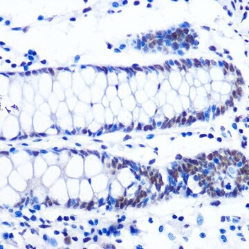
Immunocytochemistry Analysis: A 1:500 dilution from a representative lot detected PGC-1 alpha in A431, HUVEC, and NIH/3T3 cells.
Research Category
Epigenetics & Nuclear Function

Quality

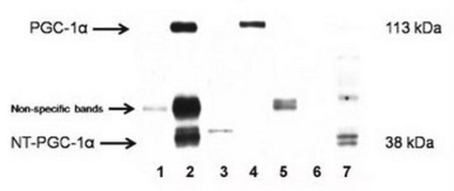
Evaluated by Western Blotting in human brown adipose tissue lysate.

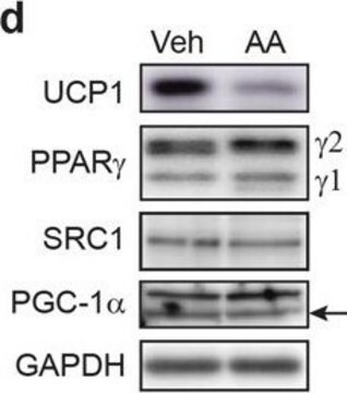
Western Blotting Analysis: 2 µg/mL of this antibody detected PGC-1 alpha in 10 µg of human brown adipose tissue lysate.

Target description

~100 kDa observed; 90.59 kDa calculated. Uncharacterized bands may be observed in some lysate(s).

Physical form

Affinity Purified
Purified rabbit polyclonal antibody in buffer containing 0.1 M Tris-Glycine (pH 7.4), 150 mM NaCl with 0.05% sodium azide.

Storage and Stability

Stable for 1 year at 2-8°C from date of receipt.

Other Notes

Concentration: Please refer to lot specific datasheet.

Disclaimer

Unless otherwise stated in our catalog or other company documentation accompanying the product(s), our products are intended for research use only and are not to be used for any other purpose, which includes but is not limited to, unauthorized commercial uses, in vitro diagnostic uses, ex vivo or in vivo therapeutic uses or any type of consumption or application to humans or animals.

Not finding the right product?  

Try our Product Selector Tool.

Storage Class Code

12 - Non Combustible Liquids

WGK

WGK 1

Flash Point(F)

does not flash

Flash Point(C)

does not flash


Certificates of Analysis (COA)

Search for Certificates of Analysis (COA) by entering the products Lot/Batch Number. Lot and Batch Numbers can be found on a product’s label following the words ‘Lot’ or ‘Batch’.

Already Own This Product?

Find documentation for the products that you have recently purchased in the Document Library.

Visit the Document Library

Zhen-Hua Wang et al.
Frontiers in pharmacology, 12, 671563-671563 (2021-06-01)
Lack of vascularization is directly associated with refractory wound healing in diabetes mellitus (DM). Enrichment of endothelial precursor cells (EPCs) is a promising but challenging approach for the treatment of diabetic wounds. Herein, we investigate the action of nicotinamide riboside
Bilal A Mir et al.
Experimental physiology, 105(8), 1326-1338 (2020-05-30)
What is the central question of this study? Do elevated levels of the stress-response protein NDRG2 protect against fasting and chronic disease in mouse skeletal muscle? What is the main finding and its importance? NDRG2 levels increased in the tibialis
Samira Mansouri et al.
Journal of immunology (Baltimore, Md. : 1950), 209(11), 2114-2132 (2022-10-20)
MPYS/STING (stimulator of IFN genes) senses cyclic dinucleotides (CDNs), generates type I IFNs, and plays a critical role in infection, inflammation, and cancer. In this study, analyzing genotype and haplotype data from the 1000 Genomes Project, we found that the
Xuan Xu et al.
Cardiovascular diagnosis and therapy, 10(3), 453-469 (2020-07-23)
Myocardial mitochondrial dysfunction is the leading cause of chronic heart failure (CHF). Increased reactive oxygen species (ROS) levels, disruption of mitochondrial biogenesis and mitochondrial Ca2+([Ca2+]m) homeostasis and reduction of the mitochondrial membrane potential (ΔΨm) cause myocardial mitochondrial dysfunction. Therefore, treating

Our team of scientists has experience in all areas of research including Life Science, Material Science, Chemical Synthesis, Chromatography, Analytical and many others.

Contact Technical Service