Skip to Content
Merck
All Photos(1)

Key Documents

ABS65

Sigma-Aldrich

Anti-CPT1A Antibody

serum, from rabbit

Synonym(s):

Carnitine O-palmitoyltransferase 1, liver isoform, CPT1-L, Carnitine O-palmitoyltransferase I, liver isoform, CPT I, CPTI-L, Carnitine palmitoyltransferase 1A

Sign Into View Organizational & Contract Pricing


About This Item

UNSPSC Code:
12352203
eCl@ss:
32160702
NACRES:
NA.41
clone:
polyclonal
application:
WB
species reactivity:
rat, mouse
technique(s):
western blot: suitable
citations:
8

biological source

rabbit

Quality Level

antibody form

serum

antibody product type

primary antibodies

clone

polyclonal

species reactivity

rat, mouse

technique(s)

western blot: suitable

isotype

IgG

NCBI accession no.

UniProt accession no.

shipped in

wet ice

target post-translational modification

unmodified

Gene Information

mouse ... Cpt1A(12894)

General description

Carnitine palmitoyltransferase 1A (CPT1A, CPT1-L, or L-CPT1) is an hepatic enzyme that, together with CPT2 and carnitine-acylcarnitine translocase, mediates the oxidation of long chain fatty acids to produce energy, in periods of fasting. CPT1A is localized in the outer mitochondrial membrane and catalyzes the addition of carnitine to long-chain fatty acids thereby enabling effective transport of these molecules to the inner mitochondrial membrane where they are metabolized. CPT1A may function as part of a complex consisting of the acyl-CoA synthetase (ACSL) and the voltage-dependent anion channel (VDAC) and is regulated by malonyl-CoA. Deficiencies in CPT1A result in hypoketotic hypoglycemia.

Immunogen

GST-tagged recombinant protein corresponding to rat CPT1A.

Application

Detect CPT1A using this Anti-CPT1A Antibody validated for use in Western Blotting.
Research Category
Signaling
Research Sub Category
Developmental Signaling

Quality

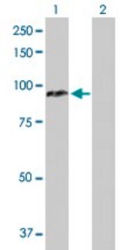
Evaluated by Western Blot in mouse liver tissue lysate.

Western Blot Analysis: A 1:1,000 dilution of this antibody detected CPT1A on 10 µg of mouse liver tissue lysate.

Target description

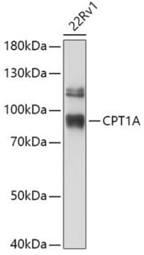
~78 kDa observed. An uncharacterized band appears at ~32 kDa in some lysates. The calculated molecular weight is 88 kDa, however CPT1A has been shown as a ~78 kDa band in western blots (Tan, L., et al (2011). J Inherit Metab Dis. 34:443–447.)

Physical form

Rabbit polyclonal serum containing 0.05% sodium azide.
Unpurified

Storage and Stability

Stable for 1 year at -20°C from date of receipt.
Handling Recommendations: Upon receipt and prior to removing the cap, centrifuge the vial and gently mix the solution. Aliquot into microcentrifuge tubes and store at -20°C. Avoid repeated freeze/thaw cycles, which may damage IgG and affect product performance.

Analysis Note

Control
Mouse liver tissue lysate

Disclaimer

Unless otherwise stated in our catalog or other company documentation accompanying the product(s), our products are intended for research use only and are not to be used for any other purpose, which includes but is not limited to, unauthorized commercial uses, in vitro diagnostic uses, ex vivo or in vivo therapeutic uses or any type of consumption or application to humans or animals.

Not finding the right product?  

Try our Product Selector Tool.

Storage Class Code

10 - Combustible liquids

WGK

WGK 1


Certificates of Analysis (COA)

Search for Certificates of Analysis (COA) by entering the products Lot/Batch Number. Lot and Batch Numbers can be found on a product’s label following the words ‘Lot’ or ‘Batch’.

Already Own This Product?

Find documentation for the products that you have recently purchased in the Document Library.

Visit the Document Library

Marion Adam et al.
FASEB journal : official publication of the Federation of American Societies for Experimental Biology, 32(6), 3434-3447 (2018-02-07)
Hydroxysteroid (17β) dehydrogenases (HSD17Bs) form an enzyme family characterized by their ability to catalyze reactions in steroid and lipid metabolism. In the present study, we characterized the phenotype of HSD17B13-knockout (HSD17B13KO) mice deficient in Hsd17b13. In these studies, hepatic steatosis
Petruta L Morvay et al.
Cell biochemistry and function, 35(3), 144-155 (2017-04-04)
The presence of peroxisomes in mammalian intestine has been revealed formerly by catalase staining combined with electron microscopy. Despite the central role of intestine in lipid uptake and the established importance of peroxisomes in different lipid-related pathways, few data are
Yi-Hsieng Samuel Wu et al.
Journal of food and drug analysis, 29(2), 375-388 (2022-06-14)
This study aims to clarify the effects of chicken liver hydrolysates (CLHs) on long-term high-fat diet (HFD)-induced insulin resistance (IR) and hepatosteatosis in mice. In vitro, the 400 μM oleic acid (OA)-added medium successfully stimulated the cellular steatosis on FL83B
Chai-Wan Kim et al.
Cell metabolism, 26(2), 394-406 (2017-08-03)
Inhibiting lipogenesis prevents hepatic steatosis in rodents with insulin resistance. To determine if reducing lipogenesis functions similarly in humans, we developed MK-4074, a liver-specific inhibitor of acetyl-CoA carboxylase (ACC1) and (ACC2), enzymes that produce malonyl-CoA for fatty acid synthesis. MK-4074
Jr-Wei Chen et al.
Poultry science, 100(4), 101009-101009 (2021-03-02)
Approximately 400 metric tons of egg chalazae, a byproduct in the liquid-egg processing plant, are produced yearly but always regarded as a waste in Taiwan. Our team successfully developed a crude egg chalaza hydrolysate by protease-A digestion (CCH-A). Free branched-chain

Our team of scientists has experience in all areas of research including Life Science, Material Science, Chemical Synthesis, Chromatography, Analytical and many others.

Contact Technical Service