Skip to Content
Merck
All Photos(2)

Key Documents

MAB1393-I

Sigma-Aldrich

Anti-PECAM-1 (CD31) Antibody, clone TLD-3A12

clone TLD-3A12, from mouse

Synonym(s):

Platelet endothelial cell adhesion molecule, CD31

Sign Into View Organizational & Contract Pricing


About This Item

UNSPSC Code:
12352203
eCl@ss:
32160702
NACRES:
NA.41
clone:
TLD-3A12, monoclonal
application:
ELISA
FACS
IHC (p)
WB
species reactivity:
human, rat
technique(s):
ELISA: suitable
flow cytometry: suitable
immunohistochemistry (formalin-fixed, paraffin-embedded sections): suitable
western blot: suitable
citations:
2

biological source

mouse

Quality Level

antibody form

purified immunoglobulin

antibody product type

primary antibodies

clone

TLD-3A12, monoclonal

species reactivity

human, rat

technique(s)

ELISA: suitable
flow cytometry: suitable
immunohistochemistry (formalin-fixed, paraffin-embedded sections): suitable
western blot: suitable

isotype

IgG1κ

NCBI accession no.

UniProt accession no.

shipped in

ambient

target post-translational modification

unmodified

Gene Information

human ... PECAM1(5175)
rat ... Pecam1(29583)

General description

Platelet endothelial cell adhesion molecule (UniProt: Q3SWT0; PECAM-1, CD31) is encoded by the Pecam1 (also known as Pecam) gene (Gene ID: 29583) in rat.PECAM-1 is a single-pass type I membrane protein that contains 6 Ig-like C2-type (immunoglobulin-like) domains. PECAM1 is differentially glycosylated involving both N-linked and O-linked glycosylation sites. It is a cell adhesion molecule that is required for leukocyte trans-endothelial migration (TEM) under most inflammatory conditions. PECAM1 plays a vital role in signaling processes involved in angiogenesis, platelet function, thrombosis, mechanosensing of endothelial cell response to fluid shear stress, and regulation of multiple stages of leukocyte migration through venular walls. PECAM1 is expressed on platelets and leukocytes and is primarily concentrated at the borders between endothelial cells. PECAM1 prevents phagocyte ingestion of closely apposed viable cells by transmitting ā€²detachmentā€² signals, and changes function on apoptosis, promoting tethering of dying cells to phagocytes. During apoptosis, the inside-out signaling of PECAM1 is disabled that allows the apoptotic cell to accept phagocyte interaction. PECAM1 is phosphorylated on serine and tyrosine residues following cellular activation. In endothelial cells Fyn is shown to mediate mechanical-force induced tyrosine phosphorylation. In response to Fc epsilon RI (FCER1 activation), PECAM1 is phosphorylated on tyrosine residues by FER and FES.

Specificity

Clone TLD-3A12 recognizes rat and human PECAM-1.

Immunogen

Lewis rat derived activated microglial cells.

Application

Immunoprecipitation Analysis: A representative lot detected PECAM-1 (CD31) in Immunoprecipitation applications (Williams, K.C., et. al. (1996). J Neurosci Res. 45(6):747-57).

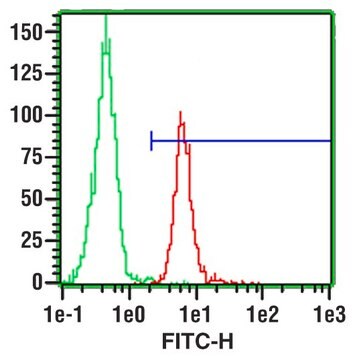
Flow Cytometry Analysis: 1 Āµg from a representative lot detected PECAM-1 (CD31) in one million rat splenocytes.

Western Blotting Analysis: A representative lot detected PECAM-1 (CD31) in Western Blotting applications (Male, D., et. al. (1995). Immunology. 84(3):453-60).

ELISA Analysis: A representative lot detected PECAM-1 (CD31) in ELISA applications (Male, D., et. al. (1995). Immunology. 84(3):453-60; Williams, K.C., et. al. (1996). J Neurosci Res. 45(6):747-57).

Affects Function: A representative lot of PECAM-1 (CD31) Affected Function (Williams, K.C., et. al. (1996). J Neurosci Res. 45(6):747-57).

Immunohistochemistry Analysis: A representative lot detected PECAM-1 (CD31) in Immunohistochemistry applications (Williams, K.C., et. al. (1996). J Neurosci Res. 45(6):747-57).
Research Category
Cell Structure
This mouse monoclonal Anti-PECAM-1 (CD31), clone TLD-3A12, Cat. No. MAB1393-I is tested for use in ELISA, Flow Cytometry, Functional Studies, Immunohistochemistry (Paraffin) and Immunohistochemistry, Immunoprecipitation, and Western Blotting, for the detection of CD31/PECAM-1.

Quality

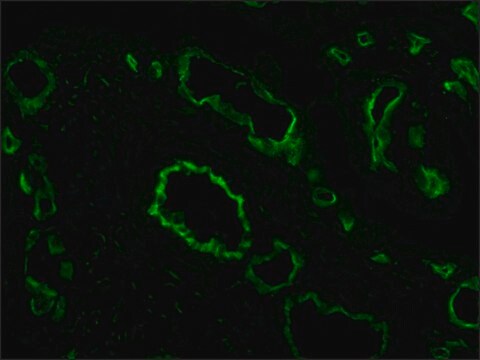
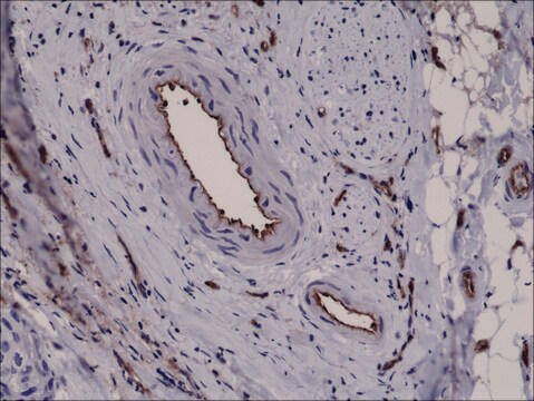
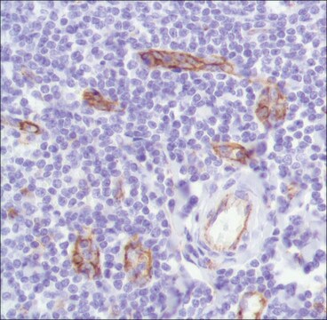
Evaluated by Immunohistochemistry in human tonsil.

Immunohistochemistry Analysis: A 1:250 dilution of this antibody detected PECAM-1 (CD31) in human tonsil tissue.

Target description

76.19 kDa calculated.

Physical form

Format: Purified
Protein G purified
Purified mouse monoclonal antibody IgG1 in PBS without azide.

Storage and Stability

Stable for 1 year at -20Ā°C from date of receipt. Handling Recommendations: Upon receipt and prior to removing the cap, centrifuge the vial and gently mix the solution. Aliquot into microcentrifuge tubes and store at -20Ā°C. Avoid repeated freeze/thaw cycles, which may damage IgG and affect product performance.

Other Notes

Concentration: Please refer to lot specific datasheet.

Disclaimer

Unless otherwise stated in our catalog or other company documentation accompanying the product(s), our products are intended for research use only and are not to be used for any other purpose, which includes but is not limited to, unauthorized commercial uses, in vitro diagnostic uses, ex vivo or in vivo therapeutic uses or any type of consumption or application to humans or animals.

Not finding the right product? Ā 

Try our Product Selector Tool.

Storage Class Code

12 - Non Combustible Liquids

WGK

WGK 2

Flash Point(F)

Not applicable

Flash Point(C)

Not applicable


Certificates of Analysis (COA)

Search for Certificates of Analysis (COA) by entering the products Lot/Batch Number. Lot and Batch Numbers can be found on a productā€™s label following the words ā€˜Lotā€™ or ā€˜Batchā€™.

Already Own This Product?

Find documentation for the products that you have recently purchased in the Document Library.

Visit the Document Library

Effect of Hypoxia-Ischemia on the Expression of Iron-Related Proteins in Neonatal Rat Brains.
Lin, et al.
Neural Plast., 2023, 4226139-4226139 (2023)
Alexander Kostyunin et al.
International journal of molecular sciences, 24(17) (2023-09-09)
Hitherto, calcified aortic valves (AVs) and failing bioprosthetic heart valves (BHVs) have been investigated by similar approaches, mostly limited to various immunostaining techniques. Having employed multiple immunostaining combinations, we demonstrated that AVs retain a well-defined cellular hierarchy even at severe

Our team of scientists has experience in all areas of research including Life Science, Material Science, Chemical Synthesis, Chromatography, Analytical and many others.

Contact Technical Service