Skip to Content
Merck
All Photos(1)

Key Documents

MAB1620

Sigma-Aldrich

Anti-Cytokeratin 5 Antibody, 6, clone D5/16B4

clone D5/16B4, Chemicon®, from mouse

Synonym(s):

58 kDa cytokeratin, Cytokeratin-5, CK-5, Keratin-5, K5, Type-II keratin Kb5

Sign Into View Organizational & Contract Pricing


About This Item

UNSPSC Code:
12352203
eCl@ss:
32160702
NACRES:
NA.41

biological source

mouse

Quality Level

antibody form

purified immunoglobulin

clone

D5/16B4, monoclonal

species reactivity

rat, mouse, human

manufacturer/tradename

Chemicon®

technique(s)

immunofluorescence: suitable
immunohistochemistry: suitable (paraffin)
western blot: suitable

isotype

IgG1

NCBI accession no.

UniProt accession no.

shipped in

wet ice

target post-translational modification

unmodified

Gene Information

human ... KRT5(3852)

General description

The cytokeratins are a family of intermediate filaments found in almost all epithelial cells. They are divided into four subclasses depending on their patterns of reactivity and electrophoretic characteristics and are also arranged I a numbered series of 20 or more acoring to molecular weight and isoelectric point (Quinlan et al. 1985). Types 5 and 6 of cytokeratin (CK5/6) are in high frequency in mesothelial cells, as opposed to cells of adenocarcinoma. CK5/6 has been shown to be a reliable marker for mesothelioma and squamous cell carcinoma of the lung and Bowen′s disease. It does not react with pulmonary adenocarcinoma.

Specificity

Expected to cross-react with mouse and rat.
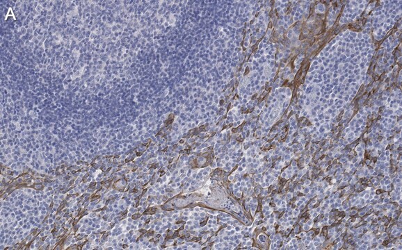
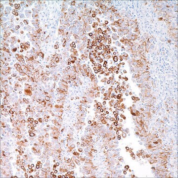
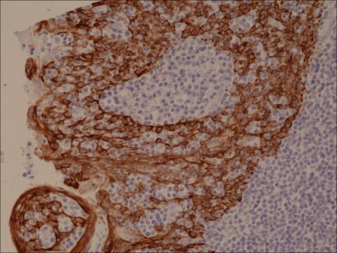
Reacts with human cytokeratin 5 on immunoblots of cytoskeletal preparations of epidermis and non-cytokeratinizing epithelium (Abstract: European Symposium of the Biology of the Cytoskeleton, Helsinki, 18-21, 6, 89, 1989) and on tissue sections. Shows reactivity with cytokeratin 6 and weak reactivity with cytokeratin 4 on immunoblots. No cross-reactivity with cytokeratins 1, 7, 8, 10, 13, 14, 18 or 19. MAB1620 recognizes basal cells and a part of the stratum spinosum in the normal pavement epithelium. The antibody may be useful for distinction of low differentiated pavement epithelium carcinoma and adenocarcinoma. The antibody works on formalin fixed tissues.

Immunogen

Purified cytokeratin 5 (Exp. Cell Res. (1986) 162:114; J. Invest. Dermatology (1987) 88:191).

Application

Anti-Cytokeratin 5 Antibody, 6, clone D5/16B4 is an antibody against Cytokeratin 5 for use in IF, IH(P) & WB.
Immunohistochemistry (frozen or paraffin sections):
A 1:50-1:100 dilution from a previous lot was used for paraffin, high-temperature antigen retrieval required (Pressure cooker, Citrate/EDTA buffer pH 6.0, 4 minutes at full pressure).

Immunofluorescence:
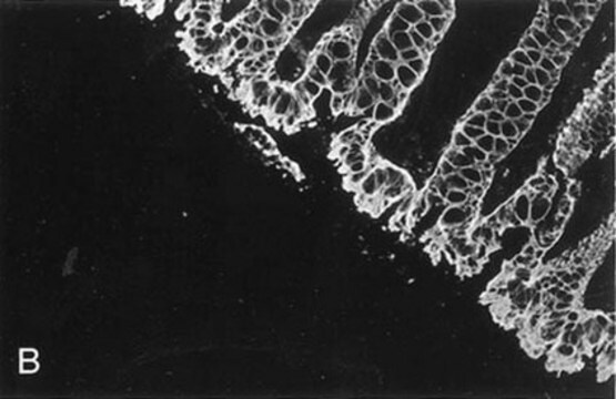
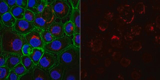
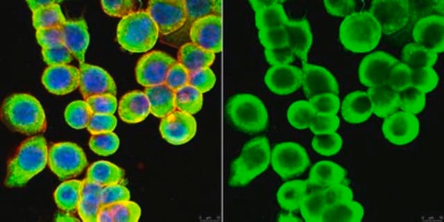
A previous lot of this antibody was used in Immunofluorescence.

Western blot:
Recognizes a 58kDa and 56kDa band corresponding to cytokeratin 5 and 6.

Optimal working dilutions must be determined by end user.
Research Category
Cell Structure
Research Sub Category
Cytoskeleton

Quality

Routinely evaluated by Western Blot on A431 lysates.

Western Blot Analysis:
1:500 dilution of this lot detected cytokeratin 5/6 on 10 ug of A431 lysates.

Target description

Cytokeratin 4 (59 kDa), Cytokeratin 5 (58 kDa) and Cytokeratin 6 (56 kDa).

Physical form

Format: Purified
Protein A purified
Purified mouse monoclonal IgG1 in buffer containing 0.02 M Phosphate buffer, 0.25 M NaCl, pH 7.6 with 0.1% sodium azide.

Storage and Stability

Stable for 6 months at 2-8°C in undiluted aliquots from date of receipt.

Analysis Note

Control
A431 cell lysate, human bladder tumor tissue.

Other Notes

Concentration: Please refer to the Certificate of Analysis for the lot-specific concentration.

Legal Information

CHEMICON is a registered trademark of Merck KGaA, Darmstadt, Germany

Disclaimer

Unless otherwise stated in our catalog or other company documentation accompanying the product(s), our products are intended for research use only and are not to be used for any other purpose, which includes but is not limited to, unauthorized commercial uses, in vitro diagnostic uses, ex vivo or in vivo therapeutic uses or any type of consumption or application to humans or animals.

Storage Class Code

10 - Combustible liquids

WGK

WGK 2


Certificates of Analysis (COA)

Search for Certificates of Analysis (COA) by entering the products Lot/Batch Number. Lot and Batch Numbers can be found on a product’s label following the words ‘Lot’ or ‘Batch’.

Already Own This Product?

Find documentation for the products that you have recently purchased in the Document Library.

Visit the Document Library

Suzete de Silva Rudland et al.
The American journal of pathology, 179(2), 1061-1072 (2011-08-02)
Two subgroups of invasive breast carcinomas have been identified with a poor prognosis in different patient cohorts: the basal-like category and the subgroup containing proteins capable of inducing metastasis in experimental rodents, the metastasis-inducing proteins (MIPs). Here we identify by
Katarzyna Sosińska-Mielcarek et al.
Breast (Edinburgh, Scotland), 22(6), 1178-1183 (2013-09-26)
There are no clinically useful biomarkers predictive of brain metastases (BM) in breast cancer. In this study, we investigated the correlation between expression of selected proteins in the primary tumor and the risk of BM in patients with metastatic breast
Indira U Mysorekar et al.
Cell host & microbe, 5(5), 463-475 (2009-05-21)
The transitional epithelium of the bladder normally turns over slowly but upon injury undergoes rapid regeneration fueled by basal uroepithelial stem and/or early progenitor cells (USCs). Little is known about the mechanisms underlying the injury response. We investigate the mechanism
Jane Muret et al.
PloS one, 7(6), e38808-e38808 (2012-06-22)
Isolated limb perfusion with TNF-α and melphalan is used with remarkable efficiency to treat unresectable limb sarcomas. Here we tested the ability of TNF-α to directly induce apoptosis of sarcoma cells. In addition, we investigated the impact of p53 in
D Mischke et al.
The Journal of investigative dermatology, 88(2), 191-197 (1987-02-01)
Human epidermal keratins from many different individuals were identified and compared by both high-resolution 1- and 2-dimensional gel electrophoresis and immunoblotting. While the polypeptide patterns obtained for keratin-enriched cytoskeletal preparations could be considered typical of normal interfollicular epidermis, they also

Our team of scientists has experience in all areas of research including Life Science, Material Science, Chemical Synthesis, Chromatography, Analytical and many others.

Contact Technical Service